
Description
PrymaLab · Research Use Only
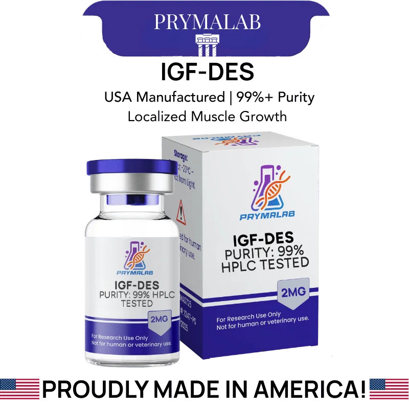
IGF-DES 2mg
des(1-3)IGF-1 · 67 residues · CAS 112603-35-7 · Reduced IGFBP affinity
IGF-1 DES 2mg supplies des(1-3)IGF-1, a 67-residue truncated analog of insulin-like growth factor 1 in which the N-terminal tripeptide Gly-Pro-Glu has been removed. That deletion substantially reduces affinity for the insulin-like growth factor binding proteins while preserving binding at the type 1 IGF receptor.
Specification Table
| Property | Value |
|---|---|
| Compound | des(1-3)IGF-1 |
| Alternative names | IGF-1 DES, IGF-DES, destripeptide IGF-1 |
| CAS number | 112603-35-7 |
| Molecular formula | C319H501N91O96S7 |
| Molecular weight | 7371.48 g/mol |
| Residue count | 67 amino acids |
| Parent molecule | Human IGF-1 (70 residues) |
| Structural modification | N-terminal Gly-Pro-Glu tripeptide removed |
| Primary molecular target | Type 1 IGF receptor (IGF-1R) |
| Binding protein affinity | Substantially reduced across the IGFBP family |
| Reported potency, L6 myoblast assay | Half-maximal protein synthesis at 1.5 ng/ml, against 13 ng/ml for intact IGF-1 |
| Reported potency ratio | Approximately 10-fold over IGF-1 in cultured cell systems |
| Disulfide bonds | Three, conserved from the parent molecule |
| Vial content | 2 mg lyophilized powder |
| Appearance | White lyophilized cake |
| Purity | Per lot-specific certificate of analysis |
| Solubility | Soluble in dilute acetic acid and in bacteriostatic water |
| Storage, lyophilized | -20°C, protected from light and moisture |
| Storage, reconstituted | 2-8°C, protected from light |
| Reconstituted stability | Not established under arbitrary conditions |
| Regulatory status | No approved human or veterinary formulation in any jurisdiction |
What Does Removing Three Residues Accomplish?
Intact human IGF-1 is 70 residues. Removing the first three, Gly-Pro-Glu, produces a 67-residue molecule with a substantially different pharmacological character despite a change of under five percent in mass.
The reason is that those three residues sit at the interface where IGF-1 contacts its binding proteins. Circulating IGF-1 is almost entirely sequestered: the great majority is held in ternary complexes with IGFBP-3 and the acid-labile subunit, which regulates how much free growth factor is available to receptors at any moment. That sequestration is a control mechanism, not an inconvenience.
Truncating the N-terminal tripeptide disrupts the binding-protein interface while leaving the receptor-binding face intact. The molecule still engages the type 1 IGF receptor. It is no longer efficiently captured by the proteins that would otherwise hold it in reserve.
Bagley and colleagues established the functional significance of this region directly (Bagley et al., Biochemical Journal, 1989), and the finding has held up as the standard explanation for the potency difference.
How Large Is the Potency Difference for IGF-1 DES 2mg?
The most useful published figure comes from a protein synthesis bioassay in rat L6 myoblasts. Half-maximal stimulation required 1.5 ng/ml of des(1-3)IGF-1 against 13 ng/ml for intact IGF-1, roughly an order of magnitude apart.
Ballard, Francis and colleagues extended the comparison into an animal model, reporting enhanced potency of the truncated form relative to IGF-1 in lit/lit mice (Ballard et al., Journal of Endocrinology, 1990). That strain carries a growth hormone-releasing hormone receptor defect, which makes it a useful background for isolating IGF effects from upstream growth hormone signalling.
The mechanism behind the difference matters more than the ratio. The truncated molecule is not intrinsically better at activating the receptor. It is better at reaching the receptor, because less of it is captured en route. In a cell culture system containing binding proteins secreted by the cells themselves, that difference is large. In a system with no binding proteins present, it should largely disappear, and that prediction is a useful internal control for anyone validating a new assay.
How Does It Differ From IGF-1 LR3?
Both analogs solve the same problem by different structural means, and confusing them is common.
IGF-1 LR3 is 83 residues. It carries an arginine substitution at position 3 and a 13-residue N-terminal extension, and its molecular weight is 9117.60. The modifications reduce binding-protein affinity by roughly a hundredfold while extending the circulating half-life considerably.
des(1-3)IGF-1 is 67 residues and 7371.48 daltons. It achieves reduced binding-protein affinity by deletion rather than substitution and extension, and it does not carry the same half-life extension. Reported clearance is faster.
For experimental purposes the choice depends on whether the design needs a short, sharp exposure or a sustained one. The truncated form suits localised or short-window work. LR3 suits designs needing prolonged receptor engagement. They are not interchangeable, and the molecular weights differ enough that molar normalisation between them is essential.
What Should Be Controlled For in IGF Work?
Insulin receptor cross-reactivity is the first consideration. IGF-1 and insulin are structurally related, and at sufficient concentration IGF analogs engage the insulin receptor. Any metabolic readout should include a control that distinguishes IGF-1R signalling from insulin receptor signalling, otherwise the attribution is assumed rather than demonstrated.
Endogenous binding protein secretion is the second. Many cultured cell lines secrete IGFBPs into the medium, and the amount varies with cell density, passage number and serum content. Since the entire rationale for the truncated analog concerns binding-protein interaction, an uncharacterised and variable IGFBP background in the medium will produce inconsistent readings between experiments that otherwise look identical.
Serum-free or defined-medium conditions remove much of that variability. Where serum is required, batch consistency becomes a real experimental variable rather than a procurement detail.
How Is IGF Activity Actually Measured?
Mass on a balance says nothing about whether a growth factor preparation is functional. Three disulfide bonds and a folded tertiary structure stand between a correct molecular weight and a molecule that binds its receptor.
The protein synthesis bioassay is the classical functional readout, and it is the assay that produced the 1.5 versus 13 ng/ml comparison quoted earlier. Rat L6 myoblasts are incubated with the growth factor and incorporation of a labelled amino acid into protein is measured. It is slow, it is sensitive, and it measures the thing that actually matters.
Receptor phosphorylation is the faster modern alternative. Engagement of the type 1 IGF receptor triggers autophosphorylation, detectable by immunoblot or by phospho-specific ELISA within minutes. It reports binding and activation directly rather than a downstream consequence, which makes it better for confirming preparation integrity and worse for comparing biological potency.
Whichever readout is chosen, an intact IGF-1 comparator run in parallel is what makes the number interpretable. The entire pharmacological argument for the truncated analog is comparative, so a measurement without the comparator answers a different question than the one being asked.
Why Local and Systemic Behaviour Diverge Here
The reduced binding-protein affinity that defines this analog has a consequence that is easy to state and easy to overlook: it changes where the molecule can act, not only how strongly.
Native IGF-1 travelling through circulation is largely captured. That capture limits how far free growth factor spreads from its point of origin and gives tissues local control over their own IGF availability through locally secreted binding proteins.
A molecule that escapes capture does not respect that local control in the same way. In a system where binding proteins would normally confine activity, the truncated form spreads further before being cleared. Its shorter circulating persistence works in the opposite direction, limiting total exposure time.
The net behaviour depends on which effect dominates in a given system, and that is not predictable from first principles. It is measurable, and worth measuring, if the experimental question concerns spatial distribution rather than only magnitude of response.
Reconstitution and Storage in Laboratory Practice
Growth factor proteins of this size are more fragile than short synthetic peptides. Three disulfide bonds hold the tertiary structure, and reducing conditions, extremes of pH or mechanical shear will disrupt it. A denatured preparation may still show protein by mass measurement while having lost receptor binding entirely.
Dilute acetic acid is a common reconstitution medium for this class, since the protein is more soluble and more stable at mildly acidic pH than at neutral. Introduce diluent gently down the vial wall and allow the cake to dissolve without agitation. Do not vortex.
Carrier protein is worth considering. At the low working concentrations typical of IGF work, adsorptive loss to plasticware is a real effect, and a carrier such as bovine serum albumin in the diluent reduces it substantially. Aliquot before freezing, hold lyophilized material at -20°C, and record lot, diluent, concentration and date.
Published Literature
Confirmed against the publisher record or a primary index before inclusion. For the potency figures quoted above, the 1989 and 1990 papers remain the sources, and nothing published since has displaced them.
- Bagley CJ, May BL, Szabo L, McNamara PJ, Ross M, Francis GL, Ballard FJ, Wallace JC. Biochemical Journal. 1989;259(3):665-671.
- Ballard FJ, Knowles SE, Walton PE, Edson K, Owens PC, Mohler MA, Ferraiolo BL. Journal of Endocrinology. 1990;127(3):401-409.
- Francis GL, Upton FM, Ballard FJ, McNeil KA, Wallace JC. Biochemical Journal. 1988;251(1):95-103.
- Tomas FM, Knowles SE, Owens PC, Read LC, Chandler CS, Gargosky SE, Ballard FJ. Biochemical Journal. 1991;276(Pt 2):547-554.
Frequently Asked Questions
What is IGF-1 DES 2mg?
IGF-1 DES 2mg is a lyophilized vial of des(1-3)IGF-1, a 67-residue truncated form of insulin-like growth factor 1 lacking the N-terminal Gly-Pro-Glu tripeptide. CAS 112603-35-7, molecular weight 7371.48. The truncation reduces binding-protein affinity while preserving type 1 IGF receptor binding. Laboratory research use only.
Which three amino acids are missing?
Glycine, proline and glutamate, occupying positions 1 through 3 of the intact 70-residue human IGF-1 sequence. Their removal leaves 67 residues. Those three residues sit at the interface where IGF-1 contacts its binding proteins, which is why deleting them changes the pharmacology so substantially.
Why does removing three residues increase potency?
The truncated molecule is not better at activating the receptor. It is better at reaching it. Circulating IGF-1 is largely sequestered in complexes with IGFBP-3 and the acid-labile subunit. Disrupting the binding-protein interface means less of the molecule is captured en route, so more remains free to engage the receptor.
What potency figures are published?
In a rat L6 myoblast protein synthesis bioassay, half-maximal stimulation required 1.5 ng/ml of des(1-3)IGF-1 against 13 ng/ml for intact IGF-1. That is roughly a tenfold difference. Ballard and colleagues extended the comparison into lit/lit mice, reporting enhanced potency of the truncated form in that model.
How does IGF-1 DES differ from IGF-1 LR3?
IGF-1 DES is 67 residues at 7371.48 daltons, achieving reduced binding-protein affinity by deletion. IGF-1 LR3 is 83 residues at 9117.60 daltons, achieving it through an arginine substitution at position 3 plus a 13-residue N-terminal extension, and it also carries a much longer circulating half-life. They are not interchangeable.
What experimental controls does IGF work require?
Insulin receptor cross-reactivity should be controlled for, since IGF-1 and insulin are structurally related and cross-engagement occurs at sufficient concentration. Endogenous IGFBP secretion by cultured cells is the second variable, and it changes with cell density, passage number and serum batch.
Why use dilute acetic acid for reconstitution?
Growth factor proteins of this class are more soluble and more stable at mildly acidic pH than at neutral. Three disulfide bonds hold the tertiary structure, and a denatured preparation may still register as protein by mass while having lost receptor binding completely. Gentle handling matters more here than with short synthetic peptides.
Should carrier protein be added?
It is worth considering. At the low working concentrations typical of IGF experiments, adsorptive loss to plasticware is significant. A carrier such as bovine serum albumin in the diluent reduces that loss substantially. Whether it is compatible with the downstream readout should be checked before adopting it as standard.
How should the vial be stored?
Dry material at minus 20 Celsius, sealed against light and moisture. Reconstituted material belongs between 2 and 8 Celsius in the dark, divided into aliquots ahead of the first freeze. Determine reconstituted stability internally. Borrowing a figure from another supplier document is not the same as measuring it.
How is IGF activity measured functionally?
The protein synthesis bioassay in rat L6 myoblasts is the classical readout and produced the 1.5 against 13 ng/ml comparison. Receptor phosphorylation by immunoblot or phospho-specific ELISA is the faster modern alternative, reporting binding and activation directly rather than a downstream consequence.
Why run an intact IGF-1 comparator?
The entire pharmacological argument for the truncated analog is comparative. A potency measurement without the parent molecule run in parallel answers a different question than the one being asked, and cannot establish whether the preparation is behaving as the truncated form should.
Does the truncation change where the molecule acts?
It can. Native IGF-1 is largely captured in circulation, which limits how far free growth factor spreads from its origin and gives tissues local control through locally secreted binding proteins. A molecule escaping capture does not respect that control in the same way, though its shorter persistence pulls in the opposite direction.
What is the practical difference in an assay without binding proteins?
The potency advantage should largely disappear. The truncated form is not intrinsically better at activating the receptor, only better at reaching it. In a system with no binding proteins present, that advantage has nothing to act on, and this prediction makes a useful internal control when validating a new assay.
Compliance Statement
IGF-1 DES 2mg is sold exclusively for laboratory research use. It is not a drug, food, or cosmetic product, and it is not a dietary product of any kind. It is not approved by the FDA or any comparable authority for human or veterinary use, and insulin-like growth factor analogs are prohibited in competitive sport under World Anti-Doping Agency rules. This product is not intended to diagnose, treat, cure, or prevent any disease. It must not be given to humans or animals. Purchase is restricted to qualified researchers and institutions operating within applicable laws. All handling is the responsibility of the purchasing laboratory.
Other formats of IGF-DES
IGF-DES is also stocked as IGF-1 DES Nasal Spray, IGF-DES 1mg/ml preloaded 3ml pen, IGF-1 LR3 Nasal Spray, IGF1 LR3 1mg/ml preloaded 3ml pen and IGF1 LR3 1MG. Each listing states its own quantity and concentration, and the pen and vial comparison explains what changes between formats.


















19 reviews for IGF-DES 2mg