
How to Reconstitute Peptides: The Four Ways Material Is Lost Before Anyone Measures Anything
Most reconstitution guides tell you to add the water slowly and not shake the vial. Both are correct and neither explains why, which means people follow the instruction until it becomes inconvenient and then stop. The reasons are worth knowing because they generalise: peptide is lost at the air-liquid interface, onto the glass, to the wrong pH, and to a sequence that was never going to dissolve in plain water. Only one of those four is about how hard you shake.
Research-use-only disclaimer and scope note: Peptides supplied by PrymaLab are intended strictly for in-vitro and laboratory research use and are not intended for human or veterinary use. This article covers reconstitution as a bench technique, meaning dissolving lyophilised powder in a diluent for laboratory work. It contains no dosing, injection or administration guidance of any kind, and nothing here is medical advice.

TL;DR
Room-temperature vial, room-temperature diluent, and run the liquid down the inside wall of the vial rather than jetting it onto the cake. Then leave it alone. Four things destroy material here and only one involves technique. Foam is aggregation, because the air-liquid interface unfolds peptides. Adsorption onto glass and plastic removes a meaningful fraction at low concentrations, invisibly. pH lands wherever the counterion puts it if you use unbuffered water. And solubility is decided by the sequence: a peptide is least soluble at its isoelectric point, so count the charged residues before assuming water is the right diluent. Research use only.
Run the diluent down the wall. Do not aim at the powder.
Foam is aggregation, happening at the air-liquid interface.
Adsorption onto container surfaces is invisible and real at low concentrations.
Least soluble at the isoelectric point. Count the charged residues.
Unbuffered water gives you whatever pH the counterion produces.
An empty-looking vial usually is not.
Status: research use only, bench technique, no administration guidance.
Before You Pierce Anything
Two things happen before the needle goes in and both get skipped.
Let the vial reach room temperature. A vial taken straight from a refrigerator or freezer is colder than the room, and cold glass condenses atmospheric moisture on and around the stopper. Piercing a stopper with water sitting on it introduces that water into a vial whose entire value is that it does not contain any. For a lyophilised peptide, water is the reagent that starts the degradation chemistry described in the storage and stability reference.
Bring the diluent to the same temperature. Partly for the same reason and partly because cold liquid dissolves solids more slowly, and slow dissolution tempts people into agitating, which is where the real damage begins.
On diluent choice, bacteriostatic water against sterile water is covered properly in the bacteriostatic water reference and I will not repeat it here. The short version is that bacteriostatic water contains benzyl alcohol as a preservative, which is what makes it multi-use, and that benzyl alcohol is not inert with respect to peptides. That last part connects to the preservative chemistry in the pen versus vial reference.
Why Foam Is the Enemy
Everybody says do not shake. Here is what shaking does, because the mechanism is the reason the rule is not negotiable.
An air-liquid interface is a hostile place for a peptide. Water molecules pull on each other and not on air, so anything sitting at that boundary is subjected to an asymmetric environment. A peptide or protein arriving at the interface tends to reorient so its hydrophobic regions face the air and its hydrophilic regions face the water, which means unfolding. Those hydrophobic regions were buried for a reason, and once exposed they will stick to the hydrophobic regions of the next unfolded molecule they meet.
That is aggregation. It is one of the four degradation chemistries, and unlike the other three it is mostly irreversible.
Shaking a vial does one thing efficiently: it creates enormous amounts of air-liquid interface. Every bubble is surface. Visible foam is a lot of surface.
The practical consequence is the technique everyone repeats without explanation. Introduce the diluent so it runs down the inside wall of the vial, which minimises both splashing and the depth to which the stream penetrates. Then let it sit. If it needs help, roll or swirl the vial slowly so the liquid moves without breaking its own surface.
Dissolution takes as long as it takes. Some sequences go in seconds, some need several minutes, and a few need something other than water, which is the next section.
The Peptide That Goes Onto the Glass
This one gets almost no coverage in supplier guides and I think it explains a good share of the results people find inexplicable.
Peptides adsorb to surfaces. Glass, polypropylene, polystyrene, the inside of a syringe, the wall of a pipette tip. The molecule sticks to the container and leaves the solution.
Whether that matters depends entirely on the ratio between how much peptide is present and how much surface is available. At high concentrations the fraction lost to the wall is trivial. At low concentrations it is not, because the surface area has not changed while the amount of material has fallen.
Two consequences worth holding on to.
Dilute solutions lose apparent potency faster than the chemistry predicts. If a solution assays lower than expected and shows no degradation products, the peptide has not broken down. It has left, and it is on the glass.
Serial dilution compounds it. Each transfer into a fresh vessel presents a fresh surface, and each fresh surface takes its share. A concentration-response curve built by serial dilution can end up with the low-concentration points systematically below their nominal values, and the shape of the curve distorts rather than shifting.
The standard mitigations are a carrier protein such as bovine serum albumin, or a very low concentration of a non-ionic surfactant, both of which occupy the surface so the peptide does not have to. Whether either is acceptable depends on the assay, and a carrier protein is obviously a problem if the readout involves protein.
I have no PrymaLab-specific data on the magnitude of this for any given compound and I am not going to invent a percentage. What I can say is that it is a real and well-described phenomenon in peptide handling, and that it is invisible, which is what makes it worth naming.
Solubility Is Decided by the Sequence
The most common support email I get is some version of "it will not dissolve, is the product bad." The answer is almost always no, and the reason follows the same principle as everything else in this library: the sequence decides.
A peptide carries charge from its ionisable side chains. The isoelectric point, written pI, is the pH at which those charges cancel and the molecule has no net charge. And a molecule with no net charge has nothing pushing it away from its neighbours, so a peptide is least soluble at its pI. That is where it precipitates.
Which gives a rule you can apply from the sequence alone, without a calculation.
Count the acidic residues, aspartate and glutamate, against the basic residues, lysine, arginine and histidine.
- Net basic, meaning more K, R and H than D and E: the peptide is positively charged at low pH, so it generally dissolves in dilute acetic acid. Then dilute into your working buffer.
- Net acidic, meaning more D and E: negatively charged at high pH, so it generally goes into dilute ammonium hydroxide or a mild base first.
- Roughly balanced or strongly hydrophobic: water will fight you. A small volume of an organic solvent such as DMSO or acetonitrile to wet the material first, then dilution into aqueous, is the usual route.
Run that against compounds in this catalogue and the answers differ. BPC-157 is GEPPPGKPADDAGLV: one lysine against two aspartates and one glutamate, so net acidic. KPV is Lys-Pro-Val, a single basic residue and nothing acidic, so strongly net basic and easy in dilute acid. Sequences heavy in leucine, isoleucine, valine and phenylalanine with few charged residues are the ones that refuse water entirely.
What pH You Actually End Up At
Unbuffered water has essentially no capacity to resist a pH change, which means that when you dissolve a peptide salt in it, the resulting pH is set by whatever you dissolved.
Synthetic peptides come off purification as salts, most commonly acetate or trifluoroacetate. Dissolve a TFA salt in pure water and the solution goes acidic, because trifluoroacetic acid is strong. Acetate is milder and the resulting pH is closer to neutral, though still on the acidic side.
Two reasons that matters rather than being trivia.
The degradation chemistry is pH-dependent. Aspartyl hydrolysis accelerates as pH falls, which is the acid-lability point made in the BPC-157 reference. Deamidation runs faster at higher pH. So the pH your solution lands at determines which of the two clocks is running faster.
The counterion is not stated on most certificates. If you do not know which salt you have, you cannot predict the pH you will get, and the same nominal reconstitution produces different solutions from different suppliers. That is one of several reasons the counterion question is worth pushing on.
Blends Are Harder, and Nobody Says So
Everything above assumed one peptide in the vial. A blend contains several, and the solubility rule from two sections up creates a problem that single-compound guides cannot warn you about.
Each peptide in a blend has its own isoelectric point, set by its own charged residues. You get one diluent for all of them. So whichever component sits closest to its pI in whatever pH you end up at is the one that comes out of solution, and it does so while the others stay dissolved.
Take the KLOW blend, which is GHK-Cu, BPC-157, TB-500 and KPV, as set out in the KLOW reference. Those four are not close to each other in charge. BPC-157 is GEPPPGKPADDAGLV, one lysine against three acidic residues, so net acidic. KPV is Lys-Pro-Val, one basic residue and nothing acidic, so strongly net basic. GHK is Gly-His-Lys, also net basic and carrying a metal ion on top.
A diluent chosen to suit the net-basic components pushes the net-acidic one toward its pI. A diluent chosen the other way does the reverse.
I do not have a clean answer to this and I would rather say so than pretend. Commercial blends are formulated with an intended diluent in mind, so following the supplier's stated diluent is the sensible default, and ours are on the product pages. What I would push back on is the assumption that a blend behaves like a single peptide during reconstitution. It has more ways to go wrong, and the failure is component-specific rather than global.
The Copper Problem
One specific case that follows directly, and it is the one where the standard solubility advice actively causes damage.
GHK-Cu is not a peptide. It is a peptide-copper complex. The tripeptide glycyl-histidyl-lysine binds a copper(II) ion with very high affinity, coordinating it through the imidazole nitrogen of the histidine, the N-terminal amine, and a backbone amide nitrogen. The copper is the point. GHK without it is a different molecule with different activity.
That coordination depends on those nitrogens being available to donate electrons, and protons compete for the same nitrogens. Drop the pH far enough and the coordinating groups protonate, which destabilises the complex and releases copper.
Now apply the solubility rule naively. GHK is net basic, one histidine and one lysine with no acidic residues, so the rule says dilute acetic acid. Follow that and you have put a pH-sensitive metal complex into acid.
I am not going to give you a threshold pH, because the stability constant depends on concentration and on what else is in solution, and I have not seen a figure I would stand behind for a reconstituted research preparation. The direction is not in question: acid destabilises the complex, and the standard advice for a net-basic peptide is the wrong advice for this one.
The visible tell is colour. Intact GHK-Cu solutions are blue, from the copper coordination. A solution that is noticeably paler than expected, or that has gone from blue toward colourless, is telling you something about the complex rather than about the peptide backbone. That is a rare case in this field of a degradation signal you can see without an instrument, and it applies to GHK-Cu and to the blends containing it.
The Vial That Looks Empty
A short section because the point is small and I answer it constantly.
A few milligrams of lyophilised peptide does not look like much. Depending on how the freeze-drying ran, it can present as a proper white cake, as a thin translucent film smeared up the inside of the glass, or as almost nothing you can see at all. The cake also sometimes detaches during shipping and sits at the bottom as a fragment that moves when you tilt the vial.
People conclude the vial is empty. It very rarely is.
Judging content by eye at these quantities does not work, and the only real check is gravimetric, which requires an analytical balance and a tare weight most people do not have. In practice the answer is to reconstitute and see whether the solution behaves as expected, which is unsatisfying and true.
What the Vacuum Tells You
Lyophilised vials are usually stoppered under partial vacuum, which is why diluent gets drawn in rather than needing to be pushed.
That gives you a free quality check. If a vial takes no vacuum at all, the seal has been compromised at some point between filling and your bench. Which means atmospheric air has been in contact with the contents, and atmospheric air carries moisture, and moisture is what the lyophilisation removed.
It also raises a sterility question, which matters more for some work than others.
One vial with no vacuum could be a marginal stopper. Several from the same lot is a manufacturing observation worth reporting to whoever sold it to you.
Concentration, and the Two Things That Skew It
The arithmetic is simple. Concentration in milligrams per millilitre is the mass in the vial divided by the volume of diluent added. Two milligrams into one millilitre gives 2mg/mL. Our peptide calculator does this and the conversions around it.
Two things make the real number differ from the calculated one.
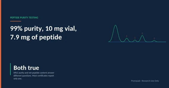
The stated mass may be the salt, not the peptide. A vial labelled 5mg may contain 5mg of peptide acetate, of which the peptide is a lower net figure, with the balance being counterion and residual water. Salt content varies by peptide and by process, and TFA salts carry proportionally more than acetate salts. Without a salt-corrected peptide content figure on the certificate, the label is an upper bound.
The solid adds volume. Dissolving 20mg of material into 1mL of water does not produce 1mL of solution, it produces slightly more. At single-digit milligram masses this is negligible. At higher masses, and particularly with the concentrated presentations, it is a real if small error in the direction of over-estimating concentration.
Neither of these matters for many purposes. Both matter if you are comparing results across suppliers or trying to reproduce someone else's conditions.
After It Is in Solution
The clock changes the moment the powder dissolves. A lyophilised peptide degrades slowly because the reactions need water. A reconstituted one has water, and hydrolysis, deamidation and aggregation all begin.
Aliquot if the material will be used more than once. Each freeze-thaw cycle is an aggregation trigger, and repeated stopper punctures introduce contamination risk and air. Dividing into single-use portions means each is thawed once.
The trade-off is the adsorption problem from earlier: smaller volumes have more surface relative to their contents, so aliquoting into many small tubes increases total adsorptive loss. If your aliquots are small and your concentration is low, you are trading one loss against another and the right answer depends on which dominates.
Protect from light if the sequence carries tryptophan, tyrosine or phenylalanine, which absorb ultraviolet and can generate species that then attack methionine and cysteine nearby. That connection is worked through in the storage reference.
Do not return unused solution to a shared vessel, and do not top up a partly used vial with fresh diluent, which produces a mixture of two ages of material at an unknown concentration.
The Failure Modes in One Place
| Failure | Mechanism | Visible? | Mitigation |
|---|---|---|---|
| Aggregation | Air-liquid interface unfolds peptide | Yes, as foam | Diluent down the wall, no shaking |
| Adsorption | Peptide sticks to glass and plastic | No | Higher concentration, carrier or surfactant |
| Precipitation | pH near the isoelectric point | Yes, as cloudiness | Match diluent to net charge |
| Hydrolysis | Acidic pH from the counterion | No | Buffer, and know your salt form |
| Moisture ingress | Condensation on a cold stopper | No | Warm to room temperature first |
| Freeze-thaw damage | Repeated ice formation | Sometimes | Aliquot to single use |
What This Article Cannot Tell You
The optimal diluent, concentration and handling for a specific sequence, because that is a property of the sequence and there are several hundred of them in this catalogue. The isoelectric point rule tells you which direction to go, not what to do.
How much material any particular compound loses to adsorption under any particular conditions. The phenomenon is well described in peptide handling generally. I have no compound-specific measurements and I am not going to estimate one.
How long a reconstituted solution of a given peptide remains usable. Establishing that requires a stability-indicating assay run over time on that material, and almost nobody in this supply chain runs one. Any number you see quoted for reconstituted shelf life is convention rather than measurement unless someone shows you the data.
What is well established: that air-liquid interfaces drive protein and peptide aggregation, that peptides adsorb to container surfaces, that solubility is minimised at the isoelectric point, and that lyophilised material is more stable than material in solution. Those are standard peptide handling and you can check them in any protein formulation text.
Frequently Asked Questions
How do you reconstitute a peptide?
Room-temperature vial and diluent, liquid introduced down the inside wall rather than onto the powder, then leave it to dissolve with gentle swirling if needed. Do not shake.
Why should you never shake a reconstituted peptide?
Shaking creates air-liquid interface, where peptides unfold and expose hydrophobic regions that then stick together. That is aggregation and it is largely irreversible. Foam settling does not undo it.
My peptide will not dissolve. Is it bad product?
Usually not. A peptide is least soluble at its isoelectric point. Count acidic residues against basic ones: net basic sequences generally go into dilute acid, net acidic into mild base, hydrophobic ones need organic solvent first.
Where does peptide go when potency drops with no visible degradation?
Onto the container. Adsorption to glass and plastic removes a meaningful fraction at low concentrations, and it is a physical loss rather than a chemical one.
Why does the diluent get pulled into the vial?
Lyophilised vials are sealed under partial vacuum. A vial with no vacuum has had a compromised seal, which raises both moisture and sterility questions.
Why is a blend harder to reconstitute than a single peptide?
Each component has its own isoelectric point and you only get one diluent. Whichever component sits nearest its pI at the resulting pH is the one that precipitates while the others stay dissolved, so the failure is component-specific rather than global.
Does GHK-Cu need different handling?
Yes. It is a peptide-copper complex, and protons compete with copper for the same coordinating nitrogens, so acid destabilises it. The standard dilute-acid advice for a net-basic peptide is the wrong advice here. Loss of the blue colour is a visible signal that the complex has been affected.
The vial looks empty. Is the peptide there?
Almost always. A few milligrams can present as a thin film on the glass. Judging by eye is unreliable at these quantities.
What concentration do you get?
Mass divided by volume. Two complications: the stated mass may be the salt rather than the peptide, and the dissolved solid adds a small volume.
Should you aliquot after reconstitution?
If it will be used across sessions, yes, because each freeze-thaw is an aggregation trigger. The trade-off is that smaller volumes have more surface area, so adsorptive loss rises.
References
- Protein and peptide aggregation at air-liquid interfaces: mechanism and formulation implications.
- Adsorption of peptides to container surfaces and its effect on solution concentration at low loadings.
- Isoelectric point and peptide solubility: standard guidance on diluent selection by net charge.
- pH dependence of aspartyl hydrolysis and asparagine deamidation in peptides.
The handling principles described here are general peptide chemistry rather than compound-specific findings. Verify against a primary source before applying them to a particular sequence.
Final disclaimer: This article is an educational research reference covering laboratory handling technique. Peptides supplied by PrymaLab are sold and studied for laboratory research use only and are not approved by any regulatory authority for human or veterinary use. Statements have not been evaluated by the FDA. Nothing here is medical advice, administration guidance, or a treatment claim.
Reconstitution as described is a bench procedure for preparing material for in-vitro research. It is not preparation for administration to humans or animals, and nothing in this article should be read as guidance for that purpose. Always verify the legal status of any research compound in your jurisdiction before purchase or use.
Formats stocked for the compounds in this article
BAC Water: BAC Water (Bacteriostatic Water) 3ML and BAC Water (Bacteriostatic Water) 10ML. Every listing is supplied for laboratory research use with a batch certificate of analysis.