
Description
PrymaLab · Research Use Only
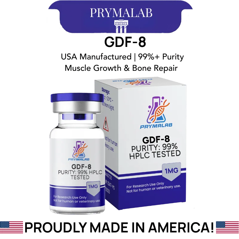
GDF-8 1mg (Myostatin)
Recombinant myostatin homodimer · TGF-β superfamily · 1mg
GDF-8 myostatin protein is a recombinant preparation of growth differentiation factor 8, a TGF-β superfamily ligand best known as a negative regulator of skeletal muscle mass. The bioactive form is a disulfide-linked homodimer of approximately 25 kDa, assembled from two monomers of roughly 12.4 kDa each.
Specification Table
| Property | Value |
|---|---|
| Compound | GDF-8 |
| Common name | Myostatin |
| Gene | MSTN |
| Protein family | Transforming growth factor beta (TGF-β) superfamily |
| Bioactive form | Disulfide-linked homodimer |
| Molecular weight, dimer | Approximately 24.8 to 25 kDa |
| Molecular weight, monomer | Approximately 12.4 kDa |
| Residue count | Approximately 109 residues per monomer in common recombinant constructs |
| Production route | Recombinant expression, commonly in E. coli |
| Glycosylation | Non-glycosylated in bacterial expression systems |
| Primary receptor | Activin receptor type IIB (ActRIIB) |
| Co-receptor | ALK4 or ALK5 type I receptor |
| Downstream signalling | Smad2 and Smad3 phosphorylation |
| Endogenous antagonists | Follistatin, the myostatin propeptide, GASP-1 and FLRG |
| Discovery | McPherron, Lawler and Lee, 1997 |
| Vial content | 1 mg |
| Appearance | White lyophilized powder |
| Purity | Per lot-specific certificate of analysis |
| Reconstitution | Commonly in dilute acid or a defined buffer per supplier instruction |
| Storage, lyophilized | -20°C or lower, protected from light and moisture |
| Storage, reconstituted | 2-8°C short term. Aliquot and freeze for longer |
| Regulatory status | Research reagent. No approved human or veterinary formulation |
This Is the Ligand, Not an Inhibitor
This distinction leads the page because it is the most consequential thing a buyer could get wrong, and the surrounding commercial language makes getting it wrong easy.
Myostatin restrains skeletal muscle growth. Compounds described as myostatin inhibitors, follistatin-based approaches, and the various agents marketed around muscle research all aim to reduce myostatin signalling. GDF-8 is myostatin. Supplying it increases the signal rather than blocking it.
That makes GDF-8 a reagent rather than an intervention. Its uses are in assay systems: as the ligand in a receptor binding study, as the target in an inhibitor screen, as a positive control establishing that a Smad reporter responds, or as the challenge in an experiment testing whether a candidate antagonist works.
McPherron, Lawler and Lee established the biology in 1997, reporting that mice lacking the gene developed dramatically increased muscle mass (McPherron et al., Nature, 1997). That paper originated the field. It defined myostatin by what its absence produces.
Anyone arriving here expecting an agent that drives muscle research endpoints has the biology inverted.
Why Does the Homodimer Matter?
GDF-8 is active only as a dimer. That single property shapes how a preparation should be handled and assessed.
Two monomers of approximately 12.4 kDa are joined by an interchain disulfide bond, giving a mature ligand near 25 kDa. TGF-β superfamily members share this architecture, and the dimeric arrangement is what allows a single ligand to bridge two receptor molecules and initiate signalling.
A monomeric preparation is inactive. That has direct analytical consequences. Non-reducing SDS-PAGE should show a band near 25 kDa, while reducing conditions collapse it to roughly 12.4 kDa. Running only reduced samples, which is the default in many laboratories, gives no information about whether the dimer formed correctly.
For bacterially expressed protein this matters more than usual. E. coli lacks the oxidising cytoplasmic environment and chaperone systems that assist disulfide formation in eukaryotic cells, so recombinant TGF-β family proteins from bacterial systems typically require in vitro refolding. Refolding yields vary between batches, and a preparation can hold correctly folded dimer alongside misfolded material at identical nominal mass.
How Does GDF-8 Myostatin Protein Signal?
Through the canonical TGF-β pathway, with activin receptor type IIB as the primary entry point.
The dimeric ligand binds two ActRIIB molecules, which then recruit type I receptors, generally ALK4 or ALK5. The type II receptor kinase phosphorylates the type I receptor, which in turn phosphorylates the receptor-regulated Smads, Smad2 and Smad3.
Phosphorylated Smad2 and Smad3 associate with Smad4 and translocate to the nucleus, where the complex regulates transcription. In muscle the net effect is restraint of growth, achieved partly through suppression of the Akt-mTOR pathway and partly through direct transcriptional effects on myogenic regulatory factors.
For assay purposes the readout follows the pathway. Smad2 or Smad3 phosphorylation by immunoblot is direct and rapid. A CAGA-luciferase reporter, which carries Smad-binding elements, gives a quantitative transcriptional readout and is the standard construct in this field.
Lee and McPherron demonstrated that follistatin and the myostatin propeptide both neutralise the ligand (Lee and McPherron, PNAS, 2001), and those antagonists remain the usual positive controls when validating an inhibition assay.
What Should an Assay Design Include?
A dimer confirmation step comes first. Non-reducing gel or size-exclusion chromatography establishes that the preparation contains the active species before any biology is attempted, and skipping it means an inactive finding cannot be distinguished from an inactive preparation.
A known antagonist provides the essential positive control. Follistatin neutralises myostatin, so a design testing a candidate inhibitor should include follistatin as a comparator establishing that the assay can detect inhibition at all.
Carrier protein deserves consideration. At the low concentrations typical of receptor work, adsorptive loss to plasticware is substantial for a protein of this size, and bovine serum albumin in the diluent reduces it. Whether it is compatible with the downstream readout should be checked first.
Finally, serum in the culture medium contains endogenous myostatin and its antagonists, which sets a background the experiment has to work against. Serum-free or defined-medium conditions remove that background, and where serum is required its batch becomes a real variable.
Why Is Myostatin Biology Unusually Well Established?
The field has a clarity most signalling biology lacks. The reason is that nature ran the experiment first.
Naturally occurring loss-of-function mutations in the myostatin gene were identified in cattle before the molecular work began, producing the double-muscled phenotype in Belgian Blue and Piedmontese breeds. Comparable mutations have since been described in dogs, sheep and a documented human case.
That matters because a knockout phenotype occurring naturally across multiple species establishes the gene function far more firmly than an engineered mouse alone. McPherron and colleagues confirmed it experimentally in 1997, and the animal genetics had already pointed the same direction.
A field with an unambiguous loss-of-function phenotype has a fixed reference point. Any proposed inhibitor can be measured against what complete absence of the protein produces, which sets an upper bound on what inhibition could achieve.
For assay work that reference point is useful in a practical way. It means the biology being measured is not in dispute, and a negative finding points to the assay rather than to uncertainty about whether the pathway matters.
How Should Activity Be Confirmed Before Use?
A recombinant protein requiring in vitro refolding should never be assumed active. Confirming it beforehand is quicker than diagnosing a failure afterward.
A Smad reporter assay is the functional check. Cells carrying a CAGA-luciferase construct respond to myostatin with measurable luminescence, and a concentration-response curve establishes both that the preparation works and what concentration range the specific lot requires.
Non-reducing gel electrophoresis is the structural check, confirming the dimer band near 25 kDa rather than a monomer at 12.4.
Running both takes an afternoon and converts an assumption into two measurements. For an inhibitor screen in particular, an inactive ligand preparation produces a screen where every candidate appears to work.
Where a lot fails either check, that is information about the lot rather than about the biology, and it should be recorded and reported to the supplier rather than worked around.
Reconstitution and Storage in Laboratory Practice
Recombinant proteins of this class are more fragile than synthetic peptides, and the folded dimeric structure is the thing being protected.
Follow the supplier reconstitution instruction rather than a generic peptide procedure. TGF-β family proteins are commonly supplied for reconstitution in dilute acid or a specific buffer, because solubility and stability are pH-dependent and the wrong medium can precipitate the protein irreversibly.
Add diluent gently and allow dissolution without agitation. Do not vortex. Mechanical shear disrupts folded structure, and a denatured preparation registers correctly by mass while having lost all activity.
Aliquot before freezing and avoid repeated freeze-thaw, which is particularly damaging to disulfide-linked dimers. Hold lyophilized material at -20°C or lower. Record lot, reconstitution medium, volume, concentration and date, and note whether dimer integrity was confirmed for the lot.
A Note on Carrier Protein
For GDF-8 myostatin protein at the low concentrations typical of receptor and reporter work, adsorptive loss for a 25 kDa protein is substantial and frequently underestimated.
Protein binds to polypropylene and glass surfaces, and the proportion lost rises as concentration falls because adsorption scales with surface area rather than with the amount in solution. At single-digit nanogram-per-millilitre concentrations the loss can dominate.
Bovine serum albumin in the diluent, typically at 0.1 percent, occupies those surfaces and reduces the loss considerably. It is standard practice for growth factors and it is often omitted for research-grade preparations.
Check compatibility with the downstream readout first. A reporter assay generally tolerates carrier protein. Some binding and mass spectrometry methods do not.
Published Literature
Each entry was checked against the publisher record or a primary index. The 1997 Nature paper founded the field and remains worth reading in full.
- McPherron AC, Lawler AM, Lee SJ. Nature. 1997;387(6628):83-90.
- Lee SJ, McPherron AC. Proceedings of the National Academy of Sciences. 2001;98(16):9306-9311.
- Thies RS, Chen T, Davies MV, Tomkinson KN, Pearson AA, Shakey QA, Wolfman NM. Growth Factors. 2001;18(4):251-259.
- Amthor H, Nicholas G, McKinnell I, Kemp CF, Sharma M, Kambadur R, Patel K. Developmental Biology. 2004;270(1):19-30.
- Rodriguez J, Vernus B, Chelh I, Cassar-Malek I, Gabillard JC, Hadj Sassi A, Seiliez I, Picard B, Bonnieu A. Cellular and Molecular Life Sciences. 2014;71(22):4361-4371.
Frequently Asked Questions
What is GDF-8 myostatin protein?
Recombinant growth differentiation factor 8, a TGF-beta superfamily ligand and negative regulator of skeletal muscle mass. The bioactive form is a disulfide-linked homodimer near 25 kDa built from two roughly 12.4 kDa monomers. Supplied at 1mg for research.
Is this a myostatin inhibitor?
No, and getting this wrong is the single most consequential error available on this page. GDF-8 is myostatin itself. Agents described as myostatin inhibitors aim to reduce this signal, whereas supplying the ligand increases it. The direction of the biology is inverted.
What is it actually used for?
Assay systems, in four common roles. As the ligand in a receptor binding study. As the target in an inhibitor screen, as a positive control establishing that a Smad reporter responds at all, or as the challenge in an experiment testing whether a candidate antagonist works.
Why does the dimer matter?
Because GDF-8 is active only as a dimer. Two monomers joined by an interchain disulfide let a single ligand bridge two receptor molecules and initiate signalling. A monomeric preparation is inactive however pure it looks by mass.
How is dimer formation confirmed?
Non-reducing SDS-PAGE should show a band near 25 kDa that collapses to roughly 12.4 kDa under reducing conditions, and running both is what makes the comparison meaningful. Reduced samples alone tell you nothing. Size-exclusion chromatography offers an alternative route to the same answer.
Why is bacterial expression relevant?
E. coli lacks the oxidising cytoplasmic environment and chaperone systems that assist disulfide formation in eukaryotic cells, so recombinant TGF-beta family proteins from bacterial systems generally require in vitro refolding. Refolding yields vary between preparations. Correctly folded dimer can coexist with misfolded material at identical nominal mass.
How does myostatin signal?
The dimeric ligand binds two activin receptor type IIB molecules, which recruit type I receptors, usually ALK4 or ALK5. The type II kinase phosphorylates the type I receptor, which phosphorylates Smad2 and Smad3, and those associate with Smad4.
What is the standard assay readout?
Smad2 or Smad3 phosphorylation by immunoblot for a direct rapid measure, or a CAGA-luciferase reporter carrying Smad-binding elements for a quantitative transcriptional readout. The CAGA construct is the standard in this field and appears throughout the literature.
What positive control should be used?
Follistatin, which neutralises myostatin and has been demonstrated to do so in published work, making it the natural comparator. Including it proves the assay can detect inhibition at all. A design testing an unknown candidate cannot otherwise demonstrate that its readout would register a hit.
How should the protein be reconstituted?
Follow the supplier instruction rather than a generic peptide procedure. TGF-beta family proteins are commonly reconstituted in dilute acid or a specific buffer, because solubility and stability are pH-dependent and the wrong medium can precipitate the protein irreversibly.
Why is myostatin biology unusually well established?
Because naturally occurring loss-of-function mutations were identified in cattle before the molecular work began, producing the double-muscled phenotype in Belgian Blue and Piedmontese breeds. Comparable mutations have since been described in dogs, sheep and a documented human case.
What does that natural evidence add?
A knockout phenotype occurring naturally across multiple species establishes gene function far more firmly than an engineered mouse alone. The animal genetics pointed the same direction as the 1997 experimental confirmation, which is a rare degree of convergence.
How does that help assay work?
It provides a fixed reference point. Any proposed inhibitor can be measured against what complete absence of the protein produces, setting an upper bound on what inhibition could achieve. A negative finding then points to the assay rather than to uncertainty about the pathway.
Compliance Statement
GDF-8 myostatin protein is sold exclusively for laboratory research use. It is not a drug, food, or cosmetic product, and it is not a dietary product of any kind. It is not approved by the FDA or any comparable authority for human or veterinary use, and it is supplied as a research reagent for in vitro assay use. This product is not intended to diagnose, treat, cure, or prevent any disease. It must not be given to humans or animals. Purchase is restricted to qualified researchers and institutions operating within applicable laws. All handling is the responsibility of the purchasing laboratory.

















19 reviews for GDF-8 1mg