Description
PrymaLab · Research use only
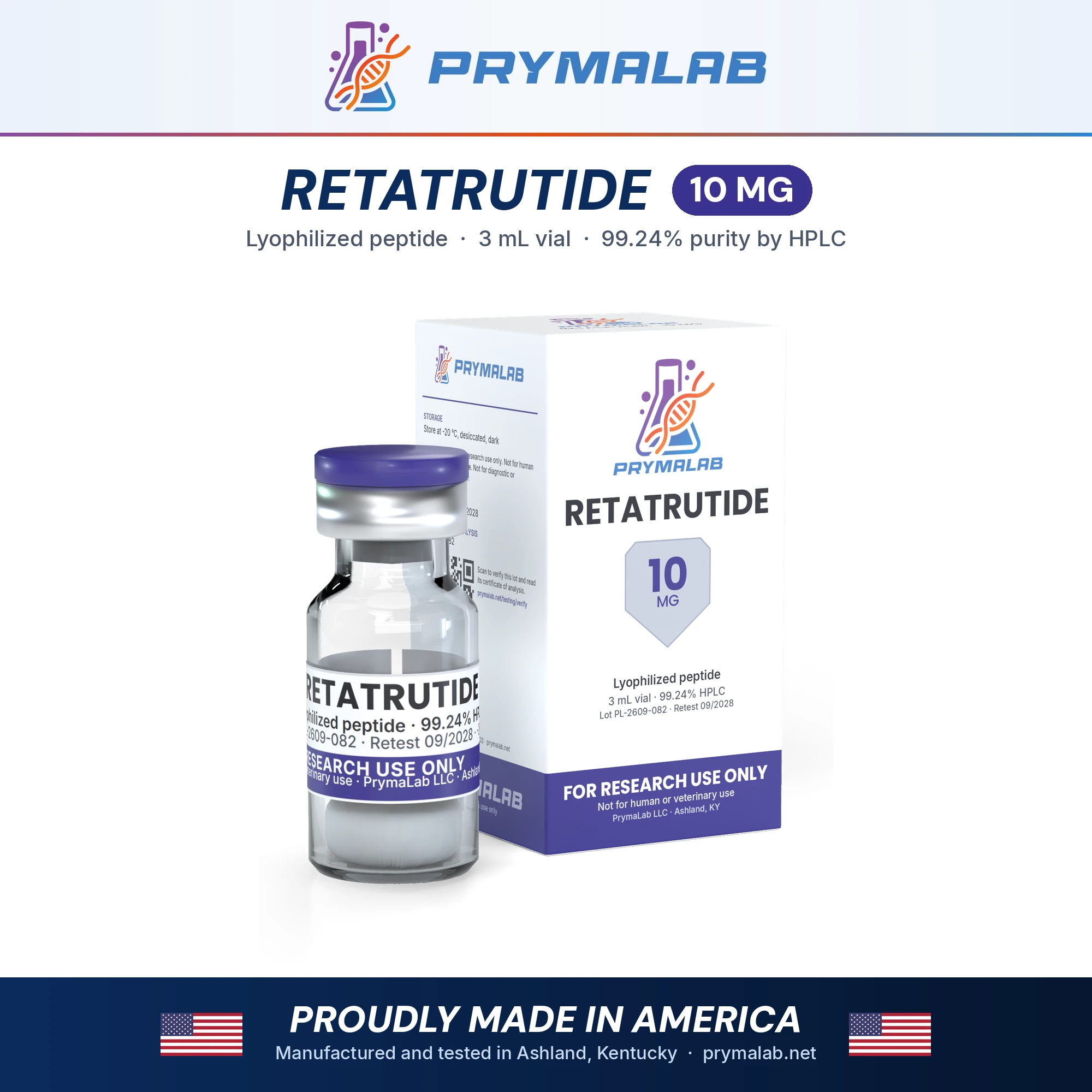
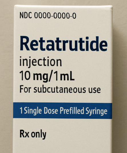
Retatrutide 10mg
LY3437943, a lipidated GIP, GLP-1 and glucagon receptor agonist, in a 10 mg vial
Retatrutide (LY3437943) is a synthetic 39-residue peptide that activates three hormone receptors at once: those for GIP, GLP-1 and glucagon. This listing is the 10 mg vial of white freeze-dried powder, the main fill in a range that runs from 5 mg to 60 mg. Published research on retatrutide moves from receptor assays and mouse studies to phase 1 and phase 2 trials in obesity, type 2 diabetes and fatty liver disease, and the compound is still investigational.
Specifications
| Property | Value |
|---|---|
| Compound | Retatrutide LY3437943; GIP/GLP-1/glucagon receptor triple agonist |
| CAS number | 2381089-83-2 |
| Molecular formula | C221H342N46O68 |
| Theoretical mass | 4731.4 Da |
| Sequence / structure | H-Y-Aib-QGTFTSDYSI-[αMeLeu]-LDKK(AEEA-γGlu-C20 diacid)AQ-Aib-AFIEYLLEGGPSSGAPPPS-NH₂ (39 residues; Aib2, α-methyl-Leu13, Aib20; Lys17 acylated with eicosanedioic acid via γ-Glu and AEEA; C-terminal amide) |
| Label claim | 10 mg/vial |
| Form | Lyophilized (freeze-dried) powder or cake in a sealed glass vial with a rubber stopper and aluminium crimp cap |
| Storage, sealed | −20 °C, desiccated, dark |
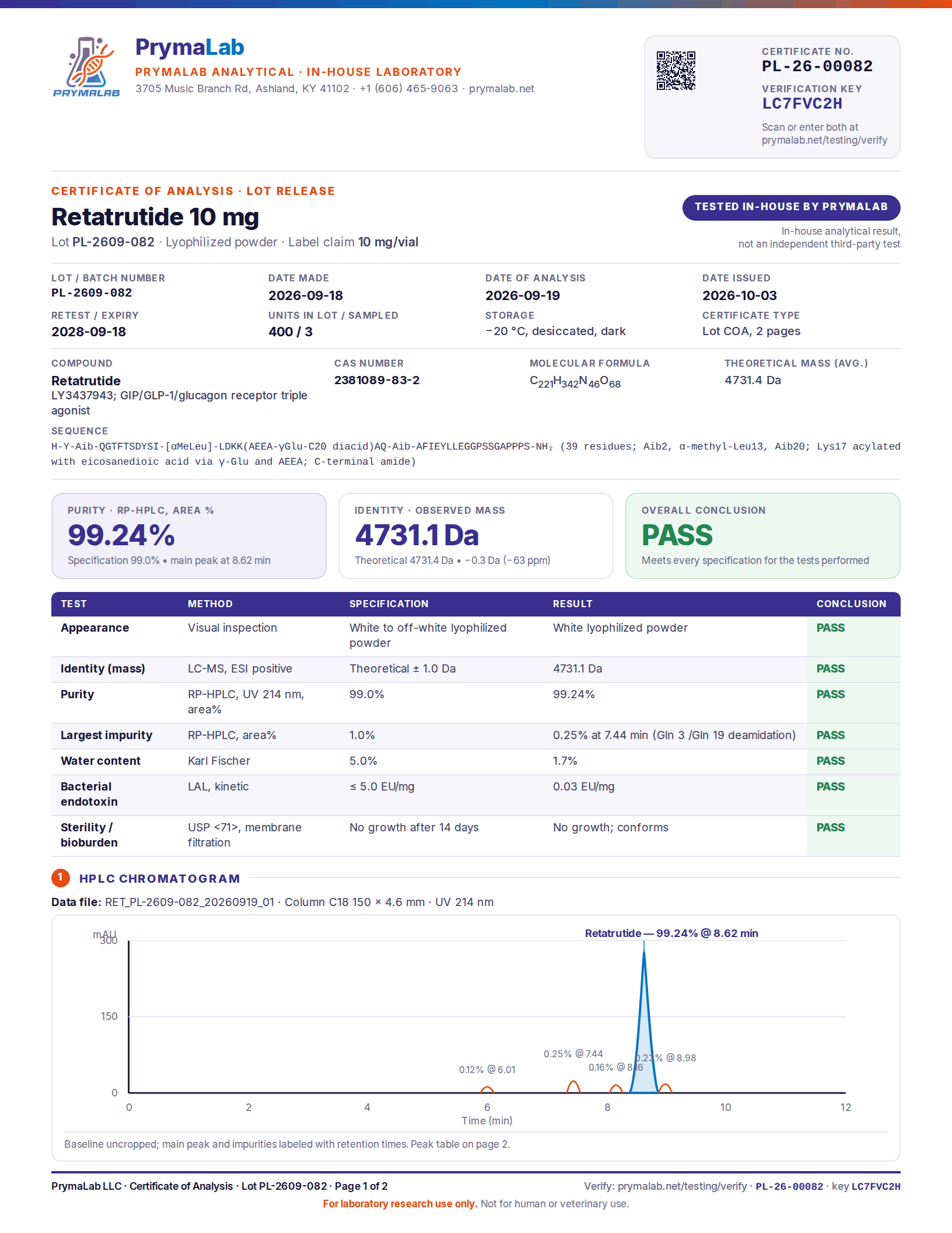
| Purity specification | 99.0% or higher by RP-HPLC |
| Lot certificate | Results for the lot now shipping are in the certificate panel on this page |
| Safety data sheet | PL-SDS-140 |
Fills in this range
Retatrutide is sold in 7 fills. The compound and the testing are the same in every fill; only the amount in the vial changes.
| Fill | Listing |
|---|---|
| 5 mg | Retatrutide 5mg |
| 10 mg | Retatrutide 10mg (this page) |
| 20 mg | Retatrutide 20mg |
| 30 mg | Retatrutide 30mg |
| 40 mg | Retatrutide 40mg |
| 50 mg | Retatrutide 50mg |
| 60 mg | Retatrutide 60mg |
Structure and molar amount
The lot certificate describes retatrutide as 39 residues with an amidated C-terminus. Three positions hold non-standard amino acids: α-aminoisobutyric acid (Aib) at positions 2 and 20 and α-methyl-leucine at position 13. Lysine 17 carries a 20-carbon diacid joined through a γ-glutamate and an AEEA linker, a lipid modification the site’s guide describes as there to bind albumin. The formula C221H342N46O68 gives a theoretical mass of 4731.4 Da, the figure the LC-MS identity result should match. At that mass 10 mg is about 2.1 µmol, slightly less in practice because the powder holds the residual water the certificate reports. Peptide purity testing explains what the certificate figures mean.
Safety data and handling
The safety data sheet classifies retatrutide as Repr. 2, H361, the GHS category for substances suspected of damaging fertility or the unborn child, and asks for the vial to be kept closed, labelled and locked up. Sealed vials belong at −20 °C, dry and dark; let one reach room temperature before piercing the stopper so moisture does not condense on the powder. Reconstituted solution goes to 2 to 8 °C, protected from light, for a period your laboratory has established, since none of the papers cited here measured retatrutide’s stability in solution. General principles are in peptide storage and stability.
What the research covers
What it is and how it acts
Retatrutide was first described under the code LY3437943 as a single peptide with agonist activity at the glucagon receptor, the glucose-dependent insulinotropic polypeptide (GIP) receptor and the glucagon-like peptide-1 (GLP-1) receptor1. In cell assays its glucagon and GLP-1 receptor activity was balanced, while its GIP receptor activity was greater1. The idea of building all three activities into one molecule is older. In 2015 Finan and colleagues reported a different monomeric triple agonist that lowered body weight, improved glycaemic control and reversed hepatic steatosis in rodent models, and confirmed each receptor’s contribution with knockout and blockade experiments2. They traced most of its effect to glucagon raising energy expenditure, GLP-1 cutting calorie intake and GIP buffering the tendency of glucagon to push blood glucose up2.
Mouse and phase 1 studies
In the retatrutide discovery paper, LY3437943 reduced body weight and improved glycaemic control in obese mice, and the authors reported that energy expenditure raised through the glucagon receptor added to the lower calorie intake driven by the GIP and GLP-1 receptors1. The same paper included a phase 1 study in which participants received the peptide once. Its safety and tolerability resembled those of other incretin drugs, and body weight was still below baseline 43 days later1.
Phase 2 trials
The phase 2 obesity trial was randomised, double-blind and placebo-controlled. It enrolled 338 adults with a BMI of 30 or more, or 27 to under 30 with at least one weight-related condition, and its primary end point was the percentage change in body weight from baseline to 24 weeks3. At 24 weeks the least-squares mean change across the retatrutide groups ranged from −7.2% to −17.5%3. A separate phase 2 trial in adults with type 2 diabetes compared retatrutide with placebo and with the GLP-1 receptor agonist dulaglutide, using a double-blind, double-dummy design across 42 US centres4.
Liver fat was the subject of a substudy within the obesity trial, in 98 participants with metabolic dysfunction-associated steatotic liver disease and at least 10% liver fat5. The mean relative change in liver fat at 24 weeks ranged from −42.9% to −82.4% in the retatrutide groups against +0.3% with placebo, and the reductions tracked changes in body weight, abdominal fat and metabolic markers of insulin sensitivity and lipid metabolism5.
What is not established
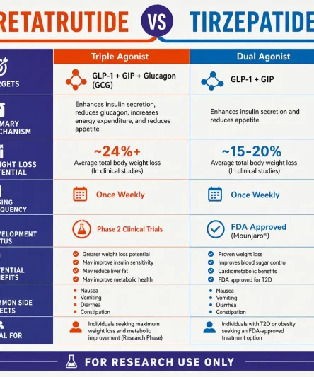
Phase 3 trials in type 2 diabetes, non-alcoholic fatty liver disease and obesity began on 28 August 20236. Their progress, including the topline announcements of 2026, is followed in the retatrutide vs tirzepatide guide, which also notes that no randomised head-to-head comparison with tirzepatide has been published. Retatrutide is not approved in any country. The human data above all come from trials of an investigational drug run under medical supervision, and the same few investigators recur across them (Frias is an author on all three phase 2 papers). How much each receptor contributes in people cannot be separated from these trial designs, since the knockout evidence for the three-receptor rationale comes from rodents2. None of the studies used PrymaLab material.
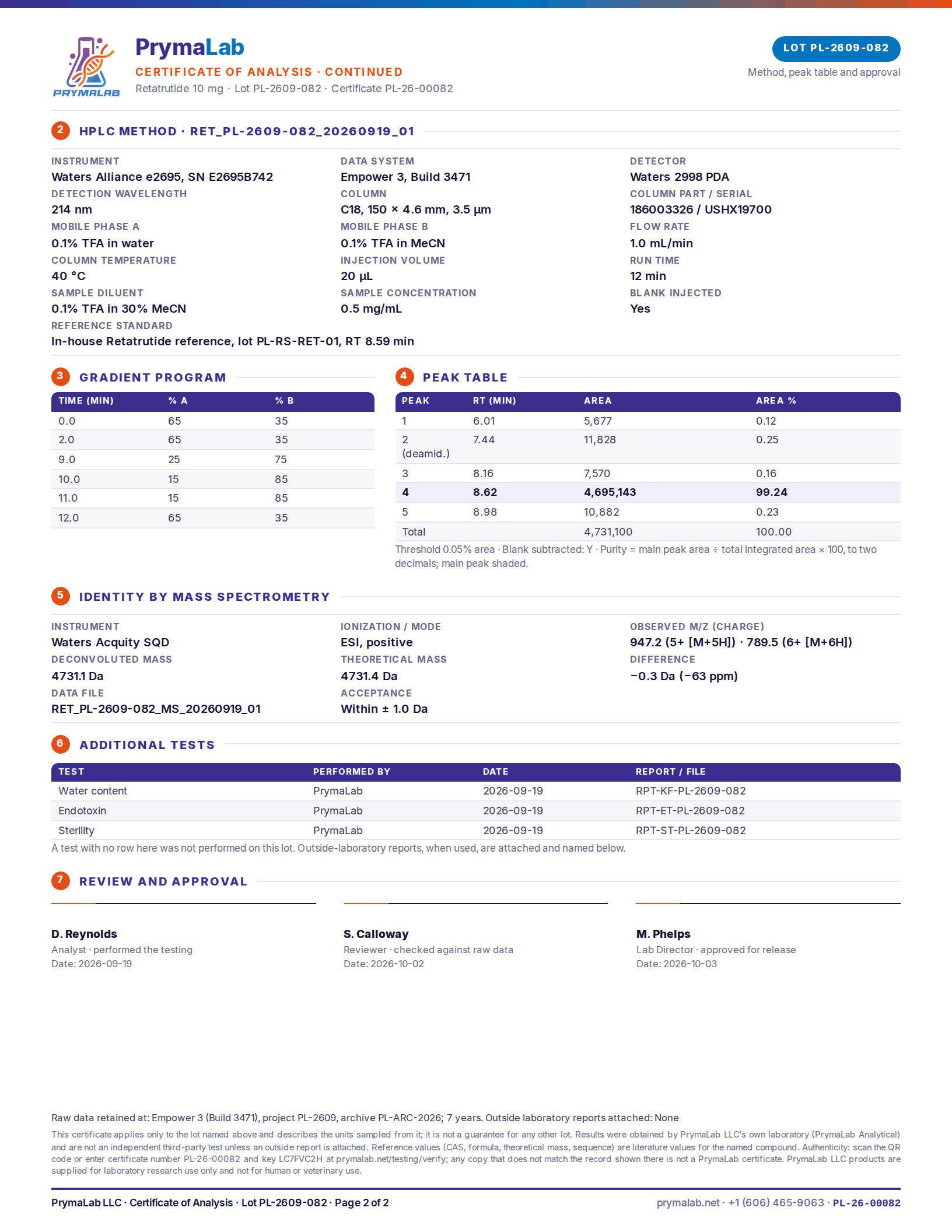
How each lot is tested
Every lot is tested in PrymaLab’s own laboratory in Ashland, Kentucky before release: RP-HPLC for purity, LC-MS for identity, Karl Fischer titration for water, LAL for endotoxin and USP <71> for sterility. The results for the lot now shipping are in the certificate panel on this page, and the certificate number, key and QR code can be checked on the verify page. These are in-house results from PrymaLab Analytical, not an independent test, and the lab is not accredited to ISO 17025. The third-party testing guarantee on this page still applies: if any independent laboratory finds the wrong compound, or less than the stated purity or amount, the order is refunded and the cost of the test is reimbursed.
Frequently asked questions
How does the 10 mg vial differ from the other retatrutide fills?
Only in the amount of powder. Every fill is the same freeze-dried retatrutide, made and tested as its own lot with its own certificate; the fills table above lists each size with the concentration it gives in 2 mL of diluent.
How do I match a certificate to the vial I received?
Read the lot number on the vial label and compare it with the certificate panel on this page, which shows the lot now shipping. Older lots are on the testing results page, and any certificate can be confirmed on the verify page with its number and key.
Which molecular weight should be used for retatrutide?
The theoretical mass on the certificate, 4731.4 Da, which follows from the formula C221H342N46O68 for the complete acylated, amidated chain. A different figure elsewhere means a different formula was assumed, so check any number against the LC-MS identity result before relying on it.
What does the research show, and what does it leave open?
Mouse studies and phase 1 and phase 2 trials measured body weight, glycaemic control and liver fat with retatrutide given under medical supervision. Phase 3 findings had appeared mostly as topline announcements by 2026, no head-to-head trial against tirzepatide exists, and none of the studies involved research-grade material.
Is retatrutide approved for human use?
No. Retatrutide is investigational, with no FDA approval, and this vial is sold for laboratory research only. It must not be given to people or animals.
References
- Coskun T, Urva S, Roell WC, et al. LY3437943, a novel triple glucagon, GIP, and GLP-1 receptor agonist for glycemic control and weight loss: From discovery to clinical proof of concept. Cell Metab. 2022;34(9):1234-1247.e9. DOI · PubMed
- Finan B, Yang B, Ottaway N, et al. A rationally designed monomeric peptide triagonist corrects obesity and diabetes in rodents. Nat Med. 2015;21(1):27-36. DOI · PubMed
- Jastreboff AM, Kaplan LM, Frías JP, et al. Triple-Hormone-Receptor Agonist Retatrutide for Obesity – A Phase 2 Trial. N Engl J Med. 2023;389(6):514-526. DOI · PubMed
- Rosenstock J, Frias J, Jastreboff AM, et al. Retatrutide, a GIP, GLP-1 and glucagon receptor agonist, for people with type 2 diabetes: a randomised, double-blind, placebo and active-controlled, parallel-group, phase 2 trial conducted in the USA. Lancet. 2023;402(10401):529-544. DOI · PubMed
- Sanyal AJ, Kaplan LM, Frias JP, et al. Triple hormone receptor agonist retatrutide for metabolic dysfunction-associated steatotic liver disease: a randomized phase 2a trial. Nat Med. 2024;30(7):2037-2048. DOI · PubMed
- Kaur M, Misra S. A review of an investigational drug retatrutide, a novel triple agonist agent for the treatment of obesity. Eur J Clin Pharmacol. 2024;80(5):669-676. DOI · PubMed
Research use only
For laboratory research use only. Retatrutide 10mg is not a drug, food, cosmetic or dietary supplement, is not approved by the FDA or any comparable authority for human or veterinary use, and must not be given to people or animals. It is sold to qualified researchers and institutions, and the buyer is responsible for lawful handling and disposal.
Other formats of Retatrutide
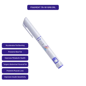
Retatrutide is also stocked as Retatrutide 15mg preloaded 3ml pen. Each listing states its own quantity and concentration, and the pen and vial comparison explains what changes between formats. The retatrutide buying guide covers what to check on a certificate before ordering.







1 review for Retatrutide 10mg