
Description
PrymaLab · Research use only
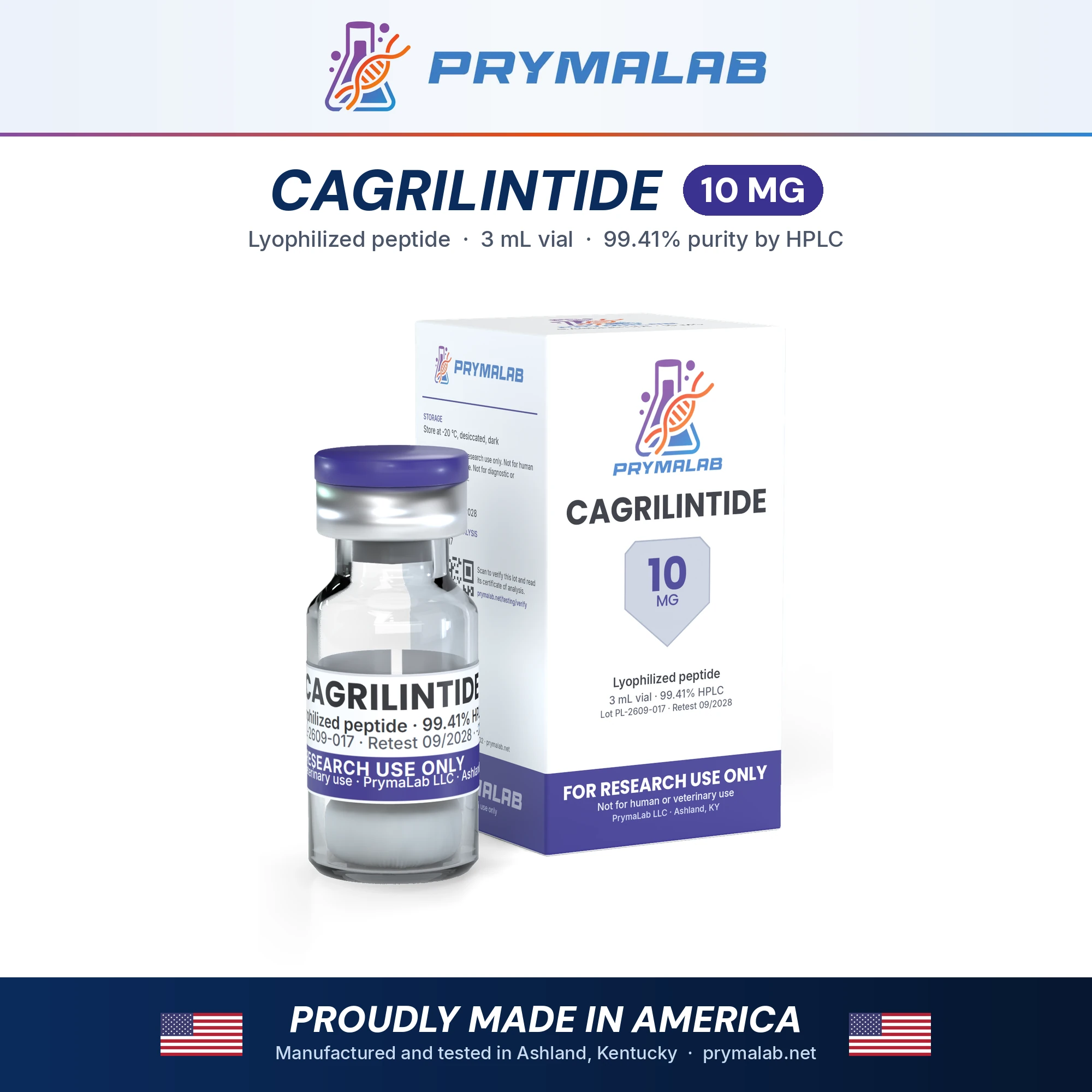
Cagrilintide 10mg
Long-acting acylated amylin analogue, 37 residues, freeze-dried in a 10 mg vial
Cagrilintide is a long-acting analogue of amylin, the 37-residue hormone that pancreatic beta cells secrete together with insulin. It keeps amylin’s disulfide and amidated C-terminus, carries prolines that curb the fibril formation human amylin is known for, and has a C20 fatty diacid on its N-terminal lysine. This listing is the 10 mg vial of freeze-dried powder, the main of two fills. Research on cagrilintide covers its design, amylin receptor pharmacology and clinical trials alone and with semaglutide.
Specifications
| Property | Value |
|---|---|
| Compound | Cagrilintide AM833; long-acting acylated amylin analogue |
| CAS number | 1415456-99-3 |
| Molecular formula | C194H312N54O59S2 |
| Theoretical mass | 4409.0 Da |
| Sequence / structure | (C20 diacid-γGlu)-KCNTATCATQRLAEFLRHSSNNFGPILPPTNVGSNTP-NH₂ (37 residues; Cys2–Cys7 disulfide; Lys1 acylated with eicosanedioic acid via γ-Glu; C-terminal amide) |
| Label claim | 10 mg/vial |
| Form | Lyophilized (freeze-dried) powder or cake in a sealed glass vial with a rubber stopper and aluminium crimp cap |
| Storage, sealed | −20 °C, desiccated, dark |
| Purity specification | 99.0% or higher by RP-HPLC |
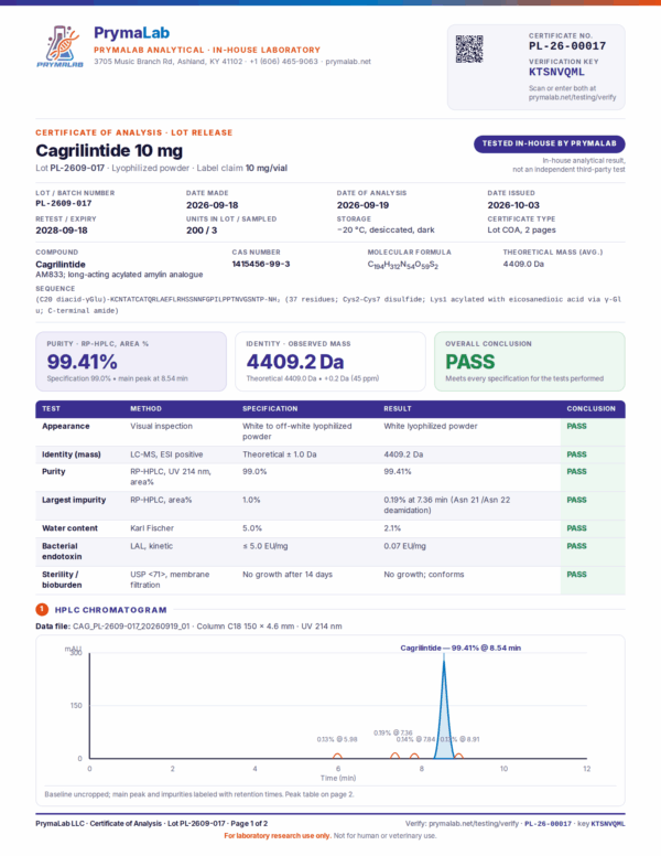
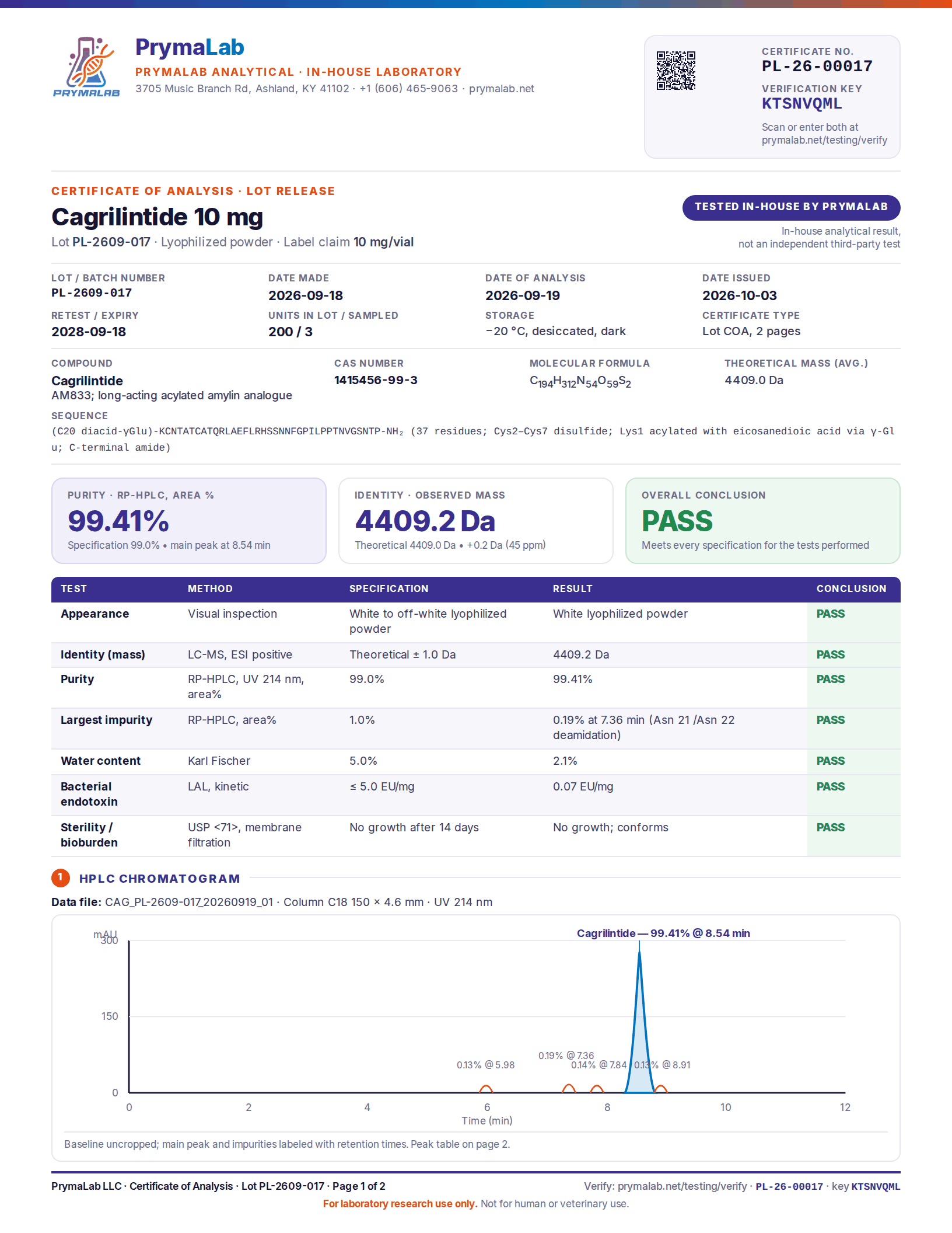
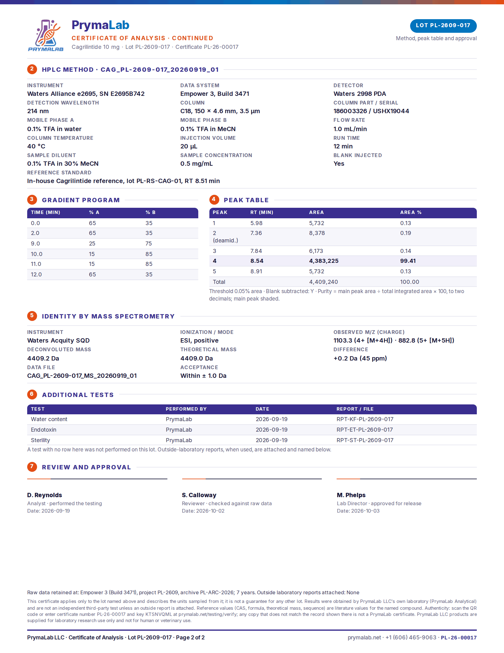
| Lot certificate | Results for the lot now shipping are in the certificate panel on this page |
| Safety data sheet | PL-SDS-026 |
Fills in this range
Cagrilintide is sold in 2 fills. The compound and the testing are the same in every fill; only the amount in the vial changes.
| Fill | Listing |
|---|---|
| 5 mg | Cagrilintide 5mg |
| 10 mg | Cagrilintide 10mg (this page) |
How cagrilintide differs from human amylin
The certificate writes cagrilintide as (C20 diacid-γGlu)-KCNTATCATQRLAEFLRHSSNNFGPILPPTNVGSNTP-NH₂. Like the native hormone it has 37 residues, a disulfide between cysteines 2 and 7 and an amidated C-terminus. Six positions are changed, as the cagrilintide and CagriSema guide sets out: glutamate 14 and arginine 17, which the developers described as a pair that stabilises the helical core, a proline at 37, and prolines at 25, 28 and 29. Human amylin has Ala-Ile-Leu-Ser-Ser at positions 25 to 29, a stretch Westermark and colleagues found strongly amyloidogenic1; cagrilintide reads Pro-Ile-Leu-Pro-Pro there.
The N-terminal lysine carries eicosanedioic acid, a C20 diacid, through a γ-glutamic acid linker, the same albumin-binding approach used for semaglutide but with a longer chain. The theoretical mass is 4409.0 Da, so 10 mg is about 2.3 µmol before allowing for the water the certificate reports, and the LC-MS identity result is the figure to compare with that mass.
What the research covers
Amylin and its aggregation problem
Amylin, also called islet amyloid polypeptide (IAPP), is one of the main secretory products of pancreatic beta cells2. Inside the islets it inhibits insulin and glucagon secretion; elsewhere it has binding sites in the brain, may contribute to satiety and slows gastric emptying2. It was discovered through its tendency to aggregate into islet amyloid, seen in type 2 diabetes in humans and in diabetic monkeys and cats, and the aggregated peptide is cytotoxic2. In 1990 Westermark and colleagues traced fibril formation to positions 20 to 29: the human and cat segments formed fibrils in vitro, three rodent segments did not, and a proline in place of serine at position 28, as in several rodents, almost completely blocked fibril formation1.
From pramlintide to a long-acting analogue
That tendency to form fibrils made amylin a hard starting point for drug design3. Pramlintide, an earlier analogue, is available for diabetes as an adjunct to insulin but has a short half-life3. Kruse and colleagues reported the structure-activity work that led to cagrilintide, compound 23 in their paper, a stable and lipidated long-acting analogue selected for clinical development with obesity as the indication3.
Receptors
Amylin acts through multi-subunit G protein-coupled receptors formed when a core receptor protein is co-expressed with receptor activity-modifying proteins (RAMPs), which gives several receptor subtypes4. In the 2015 review by Hay and colleagues, amylin acted mainly in the circumventricular organs of the central nervous system and interacted with other metabolic hormones, among them cholecystokinin, leptin and estradiol4. The core protein is the calcitonin receptor, and cagrilintide acts at the calcitonin receptor as well as at the amylin receptor complexes; the developer’s potency data are in the cagrilintide guide.
Clinical trials
A phase 2 trial at 57 sites in ten countries randomly assigned adults without diabetes, who had obesity or had overweight with hypertension or dyslipidaemia, to one of five cagrilintide strengths, liraglutide or placebo for a 26-week treatment period5. Its primary endpoint was the percentage change in body weight from baseline to week 265. A phase 1b trial at one US centre then paired cagrilintide with semaglutide in otherwise healthy adults with a BMI of 27.0 to 39.9, with treatment-emergent adverse events as the primary endpoint and pharmacokinetics among the secondary endpoints6. Phase 3a trials of the combination followed7,8; they are summarised on the Cagrilintide 5mg + Semaglutide 5mg page.
What is not established
The abstracts for the phase 2 and phase 1b trials cited here end before their results, so this page does not report them5,6. Cagrilintide is investigational, and its design history rests on one medicinal chemistry paper3. None of the cited papers measured how cagrilintide behaves in a reconstituted research vial over time, and none used PrymaLab material.
How each lot is tested
Every lot is tested in PrymaLab’s own laboratory in Ashland, Kentucky before release: RP-HPLC for purity, LC-MS for identity, Karl Fischer titration for water, LAL for endotoxin and USP <71> for sterility. The results for the lot now shipping are in the certificate panel on this page, and the certificate number, key and QR code can be checked on the verify page. These are in-house results from PrymaLab Analytical, not an independent test, and the lab is not accredited to ISO 17025. The third-party testing guarantee on this page still applies: if any independent laboratory finds the wrong compound, or less than the stated purity or amount, the order is refunded and the cost of the test is reimbursed.
Frequently asked questions
Is cagrilintide an amylin analogue or a GLP-1 agonist?
An amylin analogue. It is built on the 37-residue amylin sequence and acts at amylin receptor complexes and the calcitonin receptor, not at the GLP-1 receptor. PrymaLab also sells it combined with semaglutide, a GLP-1 analogue, in two blend fills.
How do the 5 mg and 10 mg vials differ?
Only in the amount. Both contain the same lipidated 37-residue peptide, theoretical mass 4409.0 Da, as freeze-dried powder. Each fill is made and tested as its own lot with a separate certificate, and with equal diluent the 10 mg vial gives twice the concentration.
How should the vial be stored before and after reconstitution?
Sealed, at −20 °C, desiccated and away from light; let it reach room temperature before opening. After reconstitution keep the solution at 2 to 8 °C, protected from light, and use it within the period your laboratory has established.
Is cagrilintide classed as hazardous?
Yes. The safety data sheet lists it as Repr. 2 (H361), suspected of damaging fertility or the unborn child, and asks for the vial to be kept closed, labelled and locked up, out of reach of unauthorised persons. Read the sheet before handling the powder.
Can cagrilintide be used in people or animals?
No. Cagrilintide is an investigational compound, and this vial is a research chemical rather than a drug product. It is sold for laboratory research only and must not be given to people or animals.
References
- Westermark P, Engström U, Johnson KH, et al. Islet amyloid polypeptide: pinpointing amino acid residues linked to amyloid fibril formation. Proc Natl Acad Sci U S A. 1990;87(13):5036-40. DOI · PubMed
- Westermark P, Andersson A, Westermark GT. Islet amyloid polypeptide, islet amyloid, and diabetes mellitus. Physiol Rev. 2011;91(3):795-826. DOI · PubMed
- Kruse T, Hansen JL, Dahl K, et al. Development of Cagrilintide, a Long-Acting Amylin Analogue. J Med Chem. 2021;64(15):11183-11194. DOI · PubMed
- Hay DL, Chen S, Lutz TA, et al. Amylin: Pharmacology, Physiology, and Clinical Potential. Pharmacol Rev. 2015;67(3):564-600. DOI · PubMed
- Lau DCW, Erichsen L, Francisco AM, et al. Once-weekly cagrilintide for weight management in people with overweight and obesity: a multicentre, randomised, double-blind, placebo-controlled and active-controlled, dose-finding phase 2 trial. Lancet. 2021;398(10317):2160-2172. DOI · PubMed
- Enebo LB, Berthelsen KK, Kankam M, et al. Safety, tolerability, pharmacokinetics, and pharmacodynamics of concomitant administration of multiple doses of cagrilintide with semaglutide 2·4 mg for weight management: a randomised, controlled, phase 1b trial. Lancet. 2021;397(10286):1736-1748. DOI · PubMed
- Garvey WT, Blüher M, Osorto Contreras CK, et al. Coadministered Cagrilintide and Semaglutide in Adults with Overweight or Obesity. N Engl J Med. 2025;393(7):635-647. DOI · PubMed
- Davies MJ, Bajaj HS, Broholm C, et al. Cagrilintide-Semaglutide in Adults with Overweight or Obesity and Type 2 Diabetes. N Engl J Med. 2025;393(7):648-659. DOI · PubMed
Research use only
For laboratory research use only. Cagrilintide 10mg is not a drug, food, cosmetic or dietary supplement, is not approved by the FDA or any comparable authority for human or veterinary use, and must not be given to people or animals. It is sold to qualified researchers and institutions, and the buyer is responsible for lawful handling and disposal.
Other formats of Cagrilintide
Cagrilintide is also stocked as cagrilintide 2.5mg + semaglutide 2.5mg, Cagrilintide 20mg preloaded 3ml pen, Semaglutide 5mg, Semaglutide 10mg, Semaglutide 15mg and Semaglutide 20mg. Each listing states its own quantity and concentration, and the pen and vial comparison explains what changes between formats.







Reviews
There are no reviews yet.