


Oxytocin and Neuronal Circuits: OXTR Signaling, Reward, Social Behavior, and Fear in Mammalian Research Models
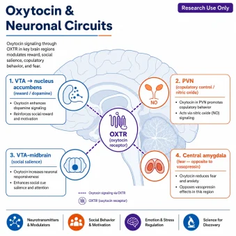
Oxytocin is a cyclic nine–amino-acid neuropeptide, traditionally known for its release from the posterior pituitary, that acts through a single receptor, the oxytocin receptor (OXTR), yet reaches an unusually wide range of neuronal circuits. This research-use-only review traces how OXTR signals inside cells and then follows oxytocin across four best-studied neural circuits in mammalian models: the mesolimbic reward system (VTA→nucleus accumbens dopamine), the PVN control of copulatory behavior, social-salience processing, and amygdala fear signaling.
Research-use-only disclaimer: Oxytocin supplied as a research chemical is intended strictly for in-vitro and laboratory research use and is not intended for human or veterinary use in that context. Every finding below is drawn from cell-culture, brain-slice, or animal models and is described in hedged, mechanistic terms. Nothing here is medical advice.

TL;DR
Oxytocin is a cyclic nonapeptide (CYIQNCPLG-NH2, ~1007 Da) that signals through the OXTR, a Gq/11-coupled GPCR driving PLC/IP3/Ca2+ plus MAPK/ERK→CREB cascades. In research models it modulates mesolimbic reward (oxytocin in the VTA raises nucleus accumbens dopamine), PVN and supraoptic nucleus (SON) copulatory circuits (via nitric oxide), social-salience processing, and amygdala fear signaling. Research use only.
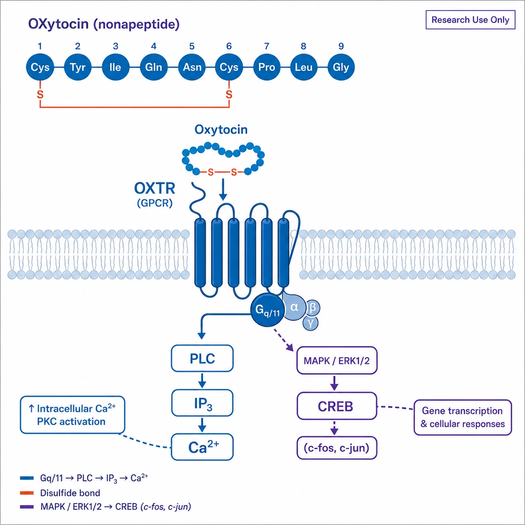
Structure: nine amino acids; disulfide ring (Cys1–Cys6) + Pro-Leu-Gly-NH2 tail; ring and amide essential for activity.
Receptor: OXTR (Gq/11) → PLC/IP3/Ca2+, plus MAPK/ERK1/2, PKC, CaMK converging on CREB (c-fos/c-jun).
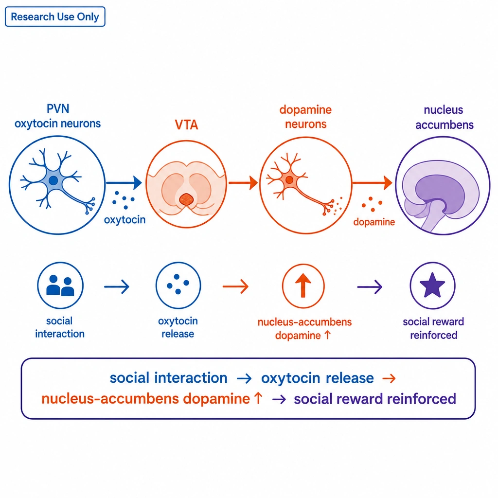
Reward: OXTR on VTA neurons projecting to NAc; oxytocin in caudal VTA raises NAc dopamine; PVN→VTA release reinforces social reward.
Copulatory PVN circuit: oxytocinergic PVN neurons drive nitric oxide (cGMP-independent) and a self-reinforcing release loop.
Fear: oxytocin and vasopressin oppositely modulate central-amygdala inputs, biasing defensive behavior.
What Is Oxytocin and How Is It Structured?
Oxytocin is a cyclic nonapeptide composed of nine amino acids in the sequence Cys-Tyr-Ile-Gln-Asn-Cys-Pro-Leu-Gly-NH2 (CYIQNCPLG-NH2), with a molecular weight of about 1007 Da. Structurally, it consists of a six-residue ring stabilized by an intramolecular disulfide bridge between the cysteines at positions 1 and 6, plus a short flexible tripeptide tail (Pro-Leu-Gly-NH2) ending in a C-terminal amide.
Both the amidated glycine and the disulfide-constrained ring are considered essential for receptor recognition: reducing the disulfide or removing the C-terminal amide greatly diminishes activity. As summarized by Gimpl & Fahrenholz (2001), most work suggests oxytocin exerts its actions by binding the OXTR in regions like the olfactory bulb, with the Pro-Leu-Gly-NH2 tail contributing selectivity over the closely related vasopressin receptors.
How Does Oxytocin Signal Through the OXTR?
The OXTR is a seven-transmembrane G-protein-coupled receptor. Its canonical route is Gq/11 coupling to phospholipase C, generating IP3 and a transient rise in intracellular Ca2+ — a link supported by radiolabeled and fluorescent binding studies. But the receptor's signaling is richer than a single cascade.
This multi-pathway signaling is why OXTR is studied not only in neurons but also in non-neuronal cells that express it, including microglia and osteoblasts — making oxytocin a probe for receptor trafficking, synaptic plasticity, calcium-dependent transcription, and MAPK-linked cell behavior across cell types. Importantly, binding studies suggest oxytocin's bioactivity is mediated almost entirely through OXTR rather than through other receptors or nonspecific membrane interactions.
How Does Oxytocin Modulate the Mesolimbic Reward System?
Oxytocin's influence on reward centers on the ventral tegmental area (VTA) and nucleus accumbens (NAc). A neuroanatomical study by Peris et al. (2017) found OXTRs on a heterogeneous population of VTA neurons projecting to the NAc, prefrontal cortex, and extended amygdala — with fewer than 10% of OXTR-expressing VTA neurons being dopaminergic (tyrosine-hydroxylase-positive). In other words, oxytocin often acts on non-dopaminergic VTA neurons that in turn shape dopamine output.
Two further studies extend this. Young et al. (2014) reported that direct oxytocin exposure to medial prefrontal cortex cells can restore pair-bonding patterns in an OXTR-dependent manner, with associated changes in NAc dopamine — evidence that oxytocin and dopamine systems interact at multiple anatomical nodes. And Chang et al. (2025) reported that VTA oxytocin signaling mediates social-isolation-induced craving for social interaction — framing the VTA as a hub where oxytocin gates social motivation.
How Does Oxytocin Control PVN Copulatory Circuits?
Oxytocin is studied as a mediator of copulatory behavior in mammalian models, and the paraventricular nucleus (PVN) is the key node. Work by Argiolas & Melis (2004) shows that oxytocinergic PVN neurons project to the posterior pituitary, extrahypothalamic, and spinal-cord targets, positioning them at the center of central copulatory control. These neurons respond to dopamine, serotonin, GABAergic, and opioid signals.
The proposed intracellular mechanism is notable: PVN oxytocinergic activation drives downstream nitric oxide (NO) production via a cGMP-independent route, and NO in turn drives oxytocin release in downstream regions — implying a self-reinforcing release loop. Complementing this, Baskerville & Douglas (2008) reported that dopamine receptors may be expressed directly on parvocellular oxytocin neurons of the PVN, making the dopamine–oxytocin axis a particularly well-defined circuit; in their words, "the PVN provides the most convincing [data] for a dopamine-oxytocin link."
How Does Oxytocin Shape Social-Salience Processing?
Beyond discrete circuits, oxytocin appears to bias how the brain weighs socially meaningful information—often involving the olfactory bulb in rodent models—a key component of social cognition and social recognition. Using functional MRI in research models, Groppe et al. (2013) reported that oxytocin better supports VTA activation in response to cues signaling either positive or negative social conditions — interpreted as OXTR engagement in midbrain and reward nodes shifting the perceived salience of social stimuli.
This salience framing has been probed in specific populations too. Greene et al. (2018) examined the effects of intranasal oxytocin on reward-circuitry responses in children with autism spectrum disorder, part of a broader research effort to understand how OXTR signaling intersects with social-reward processing. As always in this literature, these are model- and context-dependent observations, not established clinical effects.
How Does Oxytocin Modulate Amygdala Fear Signaling?
Finally, oxytocin has a well-characterized role in the amygdala. Huber et al. (2005) reported that oxytocin and vasopressin modulate excitatory inputs to neurons of the central nucleus of the amygdala (CeA) in opposite directions — providing a putative cellular mechanism by which these related neuropeptides shape fear responses.
Because CeA neurons connect to brainstem and hypothalamic neurons that organize autonomic and behavioral fear, this opposing modulation is interpreted as a candidate node where neuropeptide signaling biases the expression of defensive behavior. As reviewed by van den Burg & Hegoburu (2020), oxytocin signaling in the central amygdala may do more than simply reduce fear — it may regulate the overall style of the defensive response, adding nuance to the older "oxytocin reduces fear" picture.
| Circuit | Key node(s) | Reported effect |
|---|---|---|
| Receptor signaling | OXTR (any cell) | Gq/11 → PLC/Ca2+; MAPK/ERK → CREB |
| Mesolimbic reward | VTA → nucleus accumbens | ↑ NAc dopamine; reinforces social reward |
| Copulatory control | Paraventricular nucleus (PVN) | NO (cGMP-independent) self-reinforcing loop |
| Social salience | VTA / midbrain | Biases salience of social cues (fMRI) |
| Fear signaling | Central amygdala (CeA) | Opposite to vasopressin; shapes defense |
How is research-grade oxytocin characterized?
Because oxytocin's activity depends on its disulfide ring and C-terminal amide, identity and purity verification are essential to reproducible circuit studies. Research-grade oxytocin is typically confirmed by reversed-phase HPLC for purity and mass spectrometry for identity (including the intact disulfide and amide), handled cold, and supplied for laboratory use only. At PrymaLab, research peptides are characterized with HPLC/MS verification and independent third-party testing. No specific lot data are asserted in this general reference.
Frequently Asked Questions
What is oxytocin?
Oxytocin is a cyclic nonapeptide (CYIQNCPLG-NH2, ~1007 Da) with a disulfide-closed six-residue ring and a Pro-Leu-Gly-NH2 tail. It acts mainly through the OXTR; research material is for laboratory use only.
How does the OXTR signal?
OXTR couples mainly to Gq/11, activating PLC to raise IP3 and calcium, and also engages MAPK/ERK1/2, PKC, and CaMK cascades converging on CREB (inducing c-fos/c-jun).
How does oxytocin affect reward?
OXTRs sit on VTA neurons projecting to the nucleus accumbens; oxytocin in the caudal VTA raises NAc dopamine, and social interaction drives PVN oxytocin release into the VTA to reinforce social reward.
What is oxytocin's role in the PVN?
PVN oxytocinergic neurons project to spinal and extrahypothalamic targets and drive nitric-oxide production (cGMP-independent) in a self-reinforcing release loop studied in copulatory-behavior models.
How does oxytocin influence fear?
Huber et al. showed oxytocin and vasopressin oppositely modulate central-amygdala inputs; van den Burg & Hegoburu describe oxytocin regulating the style of defensive behavior, not just reducing fear.
Is research oxytocin approved for human use?
Material sold as a research chemical is for laboratory use only and is not intended for human or veterinary use. This article covers cellular and circuit mechanisms in research models only.
References
- Gimpl G, Fahrenholz F. The oxytocin receptor system: structure, function, and regulation. Physiol Rev. 2001;81(2):629–683. PMID:11274341
- Peris J, MacFadyen K, Smith JA, et al. Oxytocin receptors are expressed on dopamine and glutamate neurons in the mouse ventral tegmental area that project to the nucleus accumbens and other mesolimbic targets. J Comp Neurol. 2017;525(5):1094–1108. PMC6483090
- Borland JM, Grantham KN, Aiani LM, et al. Role of oxytocin in the ventral tegmental area in social reinforcement. Psychoneuroendocrinology. 2018;95:128–137. PMC6109598
- Chang HT, Cheng KH, Hung YC, Hsu KS. Oxytocin signaling in the ventral tegmental area mediates social isolation-induced craving for social interaction. J Biomed Sci. 2025;32(1):37. PMC11912778
- Young KA, Liu Y, Gobrogge KL, Wang H, Wang Z. Oxytocin reverses amphetamine-induced deficits in social bonding: evidence for an interaction with nucleus accumbens dopamine. J Neurosci. 2014;34(25):8499–8506. PMC4061391
- Argiolas A, Melis MR. The role of oxytocin and the paraventricular nucleus in the sexual behavior of male mammals. Physiol Behav. 2004;83(2):309–317. PMID:15488547
- Baskerville TA, Douglas AJ. Interactions between dopamine and oxytocin in the control of sexual behavior. Prog Brain Res. 2008;170:277–290. PMID:18655889
- Groppe SE, Gossen A, Rademacher L, et al. Oxytocin influences the processing of socially relevant cues in the ventral tegmental area of the human brain. Biol Psychiatry. 2013;74(3):172–179. PMID:23419544
- Greene RK, Spanos M, Alderman C, et al. The effects of intranasal oxytocin on reward circuitry responses in children with autism spectrum disorder. J Neurodev Disord. 2018;10(1):12. PMC5870086
- Huber D, Veinante P, Stoop R. Vasopressin and oxytocin excite distinct neuronal populations in the central amygdala. Science. 2005;308(5719):245–248. PMID:15821089
- van den Burg EH, Hegoburu C. Modulation of expression of fear by oxytocin signaling in the central amygdala: From reduction of fear to regulation of defensive behavior style. Neuropharmacology. 2020;173:108130. PMID:32389750
- Jurek B, Neumann ID. The Oxytocin Receptor: From Intracellular Signaling to Behavior. Physiol Rev. 2018;98(3):1805–1908. doi:10.1152/physrev.00031.2017
Final disclaimer: This article is an educational research reference. Oxytocin supplied as a research chemical is for laboratory research use only and is not intended for human or veterinary use in that context. Statements have not been evaluated by the FDA for the research-chemical context. Nothing here should be interpreted as medical advice.
Mechanistic descriptions are observations from cell, brain-slice, and animal models (and some human imaging) that may not generalize. Always verify the legal status of any research compound in your jurisdiction before purchase or use.