



PNC-27 and Tumor Cell Lines: How a p53-Derived Peptide Targets Membrane HDM-2 to Trigger Necrosis
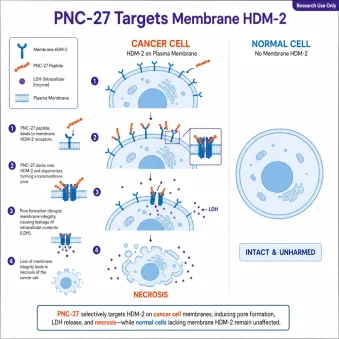
PNC-27 is a 32–amino-acid chimeric peptide that fuses a p53 fragment to a cell-penetrating leader, and it is studied for a distinctive property: killing cancer cells — but not most normal cells — by binding HDM-2 in the plasma membrane and punching pores through it. This research-use-only review examines how PNC-27 recognizes membrane HDM-2, why it drives necrosis rather than apoptosis, its newer mitochondrial actions, and its selectivity across tumor cell lines.
Research-use-only disclaimer: PNC-27 is intended strictly for in-vitro and laboratory research use. It is not a drug or therapy, is not approved for any medical use, and is not intended for human or veterinary use. All findings below come from cancer cell lines and preclinical models and are described in hedged, mechanistic terms. Nothing here is medical advice.

TL;DR
PNC-27 is a 32-residue chimeric peptide (p53 residues 12–26 + a penetratin-derived membrane residency leader) that binds HDM-2 aberrantly present in cancer-cell plasma membranes, forming a 1:1 complex and inducing transmembrane pores. The result is p53-independent necrosis (LDH release, minimal caspase/Annexin V), plus newer evidence of mitochondrial disruption. Normal cells with little membrane HDM-2 are largely spared. Research use only.
Design: chimeric peptide — p53 residues 12–26 (HDM-2-binding) fused to a penetratin-derived membrane residency peptide (MRP).
Target: HDM-2 aberrantly expressed in the plasma membrane of cancer cells; PNC-27 forms a 1:1 complex and induces pores.
Death mode: necrosis-like membrane permeabilization (LDH release) that is p53-independent — caspase-3/7 and Annexin V stay near baseline.
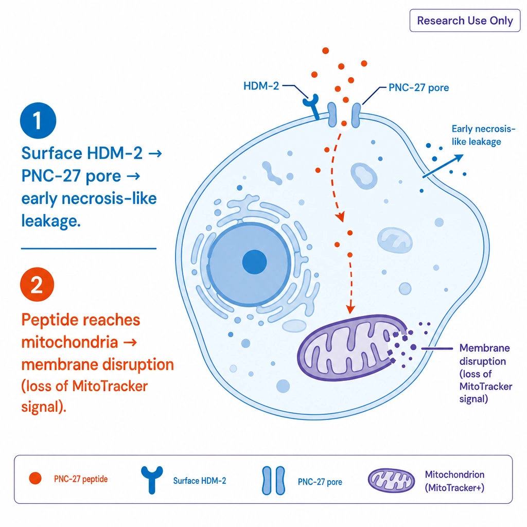
Newer finding: mitochondrial membrane disruption (loss of MitoTracker retention) supports a two-step model.
Selectivity: normal cells with low membrane HDM-2 are largely spared; caveats remain for stressed/stromal cells.
What Is PNC-27 and How Is It Built?
PNC-27 is a synthetic peptide of 32 amino acids composed of two functional segments. Its N-terminal region reproduces residues 12–26 of p53 — the portion that binds HDM-2 (the human homolog of MDM2). Its C-terminal region is a membrane residency peptide (MRP) leader derived from penetratin, a well-known cell-penetrating peptide. This makes PNC-27 a chimeric "p53-penetratin" construct, as characterized by Sarafraz-Yazdi et al. (2022).
That two-part design is the whole idea: the p53 segment provides target recognition (HDM-2), while the penetratin-derived leader controls how the peptide associates with membranes. Conformational energy calculations suggest PNC-27 adopts a p53-like binding conformation and forms 1:1 complexes with HDM-2, with the leader sequence pointing away from the complex.
How Does PNC-27 Target Cancer Cells via Membrane HDM-2?
The foundational study by Sarafraz-Yazdi et al. (2010, PNAS) established that PNC-27 kills cancer cells by binding HDM-2 that is aberrantly located in their plasma membranes and then inducing pore formation. The key selectivity signal is that significant HDM-2 appears in the membranes of many cancer cells but not in several untransformed cell lines.
Binding studies with fluorescently labeled PNC-27 show it associates with HDM-2 at the membrane in cancer cells; in HDM-2-negative membranes, the peptide diffuses inward without being retained at the surface. The causal link is compelling: when full-length HDM-2 is forced to the membrane of otherwise resistant, untransformed MCF-10-2A cells (via a CAAX membrane-targeting motif), those cells become susceptible to PNC-27-induced LDH release and loss of viability. Conversely, a membrane-localized HDM-2 variant lacking the p53/PNC-27-binding domain (residues 1–109) does not confer sensitivity — implicating the intact p53-binding pocket as the required docking site.
Necrosis vs. Apoptosis: How Does PNC-27 Kill?
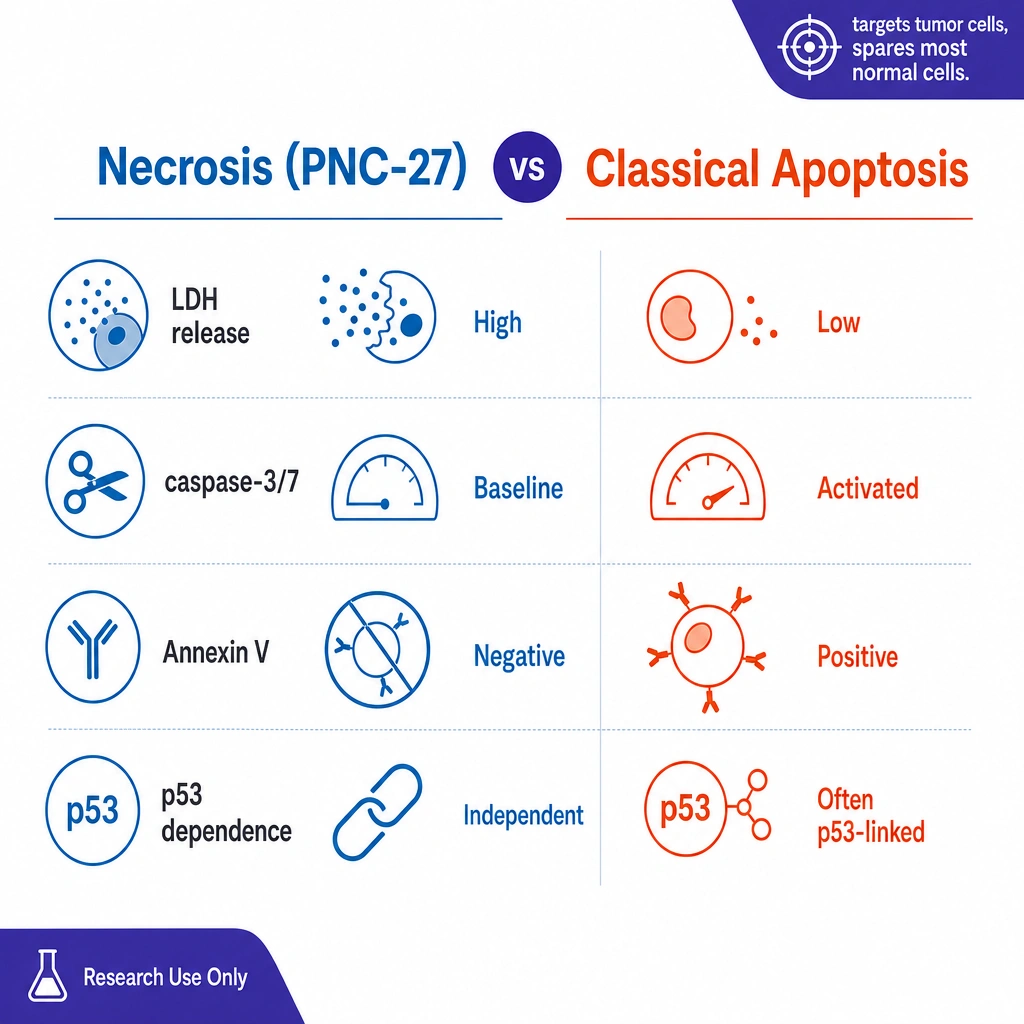
PNC-27 appears to cause a necrosis-like death driven by membrane permeabilization, not classical apoptosis. Research by Davitt et al. (2014) in the K562 leukemia model showed PNC-27 co-localizing with membrane HDM-2 by confocal microscopy, followed by LDH release and near-complete loss of viability — while caspase-3/7 activity stayed at baseline and classical apoptotic markers were absent.
Critically, this death is p53-independent: K562 cells lack p53 yet still die, so PNC-27 does not need to restore p53 signaling — it acts physically at the membrane. Similar results were reported by Thadi et al. (2020) across U937, OCI-AML3, and HL-60 leukemia lines, which express high plasma-membrane HDM-2, whereas normal mononuclear cells display only minimal surface HDM-2. After brief exposure, PNC-27 reduced viability in a concentration-dependent way with strong LDH release, while Annexin V and caspase-3 stayed near baseline.
| Readout | PNC-27 response | Classical apoptosis |
|---|---|---|
| LDH release | High (membrane leak) | Low until late |
| Caspase-3/7 activity | Near baseline | Strongly activated |
| Annexin V | Near baseline | Positive |
| p53 dependence | Independent (kills p53-null K562) | Often p53-linked |
| Primary event | Membrane pore formation | Intracellular cascade |
| Target requirement | Membrane HDM-2 | Varies |
Does PNC-27 Also Disrupt Mitochondria?
Recent work extends PNC-27's membrane activity inside the cell. Krzesaj et al. (2024) proposed that PNC-27 is "not restricted to acting at the plasma membrane via HDM-2" but also "binds to the membranes of mitochondria, resulting in their disruption" once it gains intracellular access. They studied PNC-27-exposed MIA-PaCa-2 pancreatic carcinoma cells with organelle-selective dyes and ultrastructural methods.
These observations support a two-step model: PNC-27 first targets membrane HDM-2 at the cell surface, inducing pore formation and early necrosis-like leakage; it then extends its membrane-disruptive activity to mitochondria, amplifying bioenergetic failure. Its amphipathic helix-loop-helix structure is posited to form or expand pores in mitochondrial membranes.
Which Tumor Cell Lines Respond to PNC-27?
Across the referenced studies, PNC-27 activity has been reported in a range of tumor cell types that share high membrane HDM-2:
- Leukemia lines: K562 (p53-null), U937, OCI-AML3, HL-60.
- Pancreatic carcinoma: MIA-PaCa-2 (mitochondrial-disruption studies).
- Breast models: MCF-7 (susceptible) vs. untransformed MCF-10-2A (resistant unless membrane HDM-2 is forced in).
- Additional solid tumors: the same membrane-HDM-2 mechanism has been examined in further carcinoma models in follow-up work.
The unifying variable is not tissue of origin but whether HDM-2 is present in the plasma membrane. That is why the researchers frame aberrant membrane HDM-2 as a candidate distinguishing feature between malignant and non-malignant cells in these systems.
What Are the Selectivity Caveats?
PNC-27's selectivity is real in the tested panels but not unlimited. As noted by Aguon et al. (2017) and related commentary, selectivity assays are often limited to a small set of non-tumor lines, which may underestimate heterogeneity in HDM-2 membrane expression or overlook stressed and inflamed non-tumor cells.
If PNC-27 forms pores wherever membrane HDM-2 is present, then endothelial cells, stromal cells, or regenerating epithelia that transiently express surface HDM-2 could, in principle, also be affected — a theoretical concern that motivates testing across wider tumor and non-tumor panels before any strong claims of tumor exclusivity. This is a case where the mechanism's strength (a single, physical target) is also the source of its main caveat.
How is research-grade PNC-27 characterized?
For membrane and cell-death assays, peptide identity and purity directly affect pore-formation readouts, so verification matters. Research-grade PNC-27 is typically confirmed by reversed-phase HPLC for purity and mass spectrometry for identity, handled cold, and supplied for laboratory use only. At PrymaLab, research peptides are characterized with HPLC/MS verification and independent third-party testing. No specific lot data are asserted in this general reference.
Frequently Asked Questions
What is PNC-27?
PNC-27 is a 32-amino-acid chimeric peptide combining p53 residues 12–26 (HDM-2-binding) with a penetratin-derived membrane residency leader. It binds membrane HDM-2 on cancer cells and induces necrosis, and it is for research use only.
How does PNC-27 kill cancer cells?
It binds HDM-2 in the cancer-cell plasma membrane, forms a 1:1 complex, and induces transmembrane pores that leak intracellular contents (LDH release), producing a necrosis-like death. An anti-HDM-2 (p53-site) antibody blocks this effect.
Is the death apoptosis or necrosis?
Necrosis-like. Caspase-3/7 and Annexin V remain near baseline while LDH release is high, and killing is p53-independent (K562 cells lack p53 yet still die).
Does PNC-27 affect mitochondria?
Yes, per Krzesaj et al. (2024): treated MIA-PaCa-2 cells lose MitoTracker retention while keeping LysoTracker, and the peptide localizes to mitochondrial membranes — supporting a two-step plasma-membrane-then-mitochondria model.
Why does PNC-27 spare normal cells?
Most normal cells have little surface HDM-2, so PNC-27 cannot dock and form pores; forcing membrane HDM-2 into resistant cells makes them susceptible. Selectivity caveats remain for stressed or stromal cells.
Is PNC-27 an approved cancer drug?
No. It is a research-use-only peptide studied in cell lines and preclinical models, not an approved therapy, and not intended for human or veterinary use.
References
- Sarafraz-Yazdi E, Bowne WB, Adler V, et al. Anticancer peptide PNC-27 adopts an HDM-2-binding conformation and kills cancer cells by binding to HDM-2 in their membranes. Proc Natl Acad Sci U S A. 2010;107(5):1918–1923. doi:10.1073/pnas.0909364107
- Krzesaj P, Adler V, Feinman RD, et al. Anti-Cancer Peptide PNC-27 Kills Cancer Cells by Unique Interactions with Plasma Membrane-Bound hdm-2 and with Mitochondrial Membranes Causing Mitochondrial Disruption. Ann Clin Lab Sci. 2024;54(2):137–148. PMID:38802154
- Davitt K, Babcock BD, Fenelus M, et al. The anti-cancer peptide, PNC-27, induces tumor cell necrosis of a poorly differentiated non-solid tissue human leukemia cell line that depends on expression of HDM-2 in the plasma membrane. Ann Clin Lab Sci. 2014;44(3):241–248. PMID:25117093
- Thadi A, Lewis L, Goldstein E, et al. Targeting Membrane HDM-2 by PNC-27 Induces Necrosis in Leukemia Cells But Not in Normal Hematopoietic Cells. Anticancer Res. 2020;40(9):4857–4867. PMID:32878773
- Sarafraz-Yazdi E, et al. PNC-27, a Chimeric p53-Penetratin Peptide, Binds to HDM-2 in a p53 Peptide-like Structure, Induces Selective Membrane-Pore Formation and Leads to Cancer Cell Lysis. Biomedicines. 2022;10(5):945. PMC9138867
- Aguon PM, Aasen T, Distler ES, Mallin E. Experimental PNC-27 Therapy and Massive GI Hemorrhage: A Complication or Coincidence? Am J Gastroenterol. 2017;112:S1035–S1036 (abstract 1879).
Final disclaimer: This article is an educational research reference. PNC-27 is sold and studied for laboratory research use only, is not approved by any regulatory authority, and is not intended for human or veterinary use. Statements about PNC-27 have not been evaluated by the FDA. Nothing here should be interpreted as medical advice or as a claim of anticancer efficacy in humans.
Mechanistic descriptions are observations from cancer cell lines and preclinical models; selectivity and effects may not generalize. Always verify the legal status of any research compound in your jurisdiction before purchase or use.